Deep Learning for Pancreas Segmentation: a Systematic Review

0
Sign in to get full access
Overview
- This is a systematic review of deep learning approaches for pancreas segmentation from medical imaging data.
- The paper examines the challenges in pancreas segmentation and how deep learning techniques have been used to address them.
- It covers the key architectural components, training strategies, and evaluation metrics reported in the literature.
Plain English Explanation
The pancreas is an important organ located behind the stomach that plays a crucial role in digestion and regulating blood sugar levels. Accurately segmenting or outlining the pancreas in medical images like CT or MRI scans is an important step for various medical applications, such as diagnosing and monitoring pancreatic diseases.
However, segmenting the pancreas is a difficult task due to its small size, variable shape and position, and similarity to surrounding organs. Deep learning, a type of artificial intelligence that excels at analyzing complex data, has shown promise in improving pancreas segmentation compared to traditional image processing techniques.
This systematic review examines the various deep learning approaches that have been proposed in the research literature for pancreas segmentation. It looks at the architectural design choices, training strategies, and evaluation metrics used to assess the performance of these deep learning models. The goal is to provide a comprehensive overview of the state-of-the-art in this area and identify future research directions.
Technical Explanation
The paper first discusses the key challenges in pancreas segmentation, including the organ's small size, variable shape and location, and similarity to surrounding structures like the stomach and spleen. These factors make it difficult for traditional image processing techniques to accurately delineate the pancreas boundaries.
To address these challenges, researchers have turned to deep learning models, which can learn complex visual patterns from large datasets of annotated medical images. The paper reviews the architectural components commonly used in these deep learning models, such as convolutional neural networks, encoder-decoder designs, and attention mechanisms. It also examines the various training strategies, including data augmentation, transfer learning, and multi-task learning, which have been employed to improve model performance.
In terms of evaluation, the review covers the common metrics used to assess pancreas segmentation accuracy, such as Dice coefficient, Jaccard index, and average surface distance. It also discusses how these models have been validated on publicly available datasets and in multi-center studies to ensure robustness.
Critical Analysis
The paper provides a comprehensive overview of the state-of-the-art in deep learning for pancreas segmentation, highlighting the significant progress made in this area. However, it also acknowledges several limitations and areas for further research:
- The majority of studies have focused on CT imaging, with limited work on MRI data. Expanding deep learning approaches to diverse imaging modalities is an important next step.
- Most studies use relatively small datasets from single institutions, which could limit the generalizability of the models. Larger, multi-center datasets are needed to develop more robust and clinically-applicable solutions.
- There is a lack of attention to segmentation of pathological pancreases, such as those with tumors or other abnormalities. Incorporating these cases is crucial for real-world clinical deployment.
- The review does not delve into the computational efficiency and inference speed of the deep learning models, which are important factors for practical clinical use.
Conclusion
This systematic review provides a detailed analysis of the progress made in deep learning for pancreas segmentation from medical imaging data. The findings highlight the potential of these AI-powered techniques to address the longstanding challenges in this domain. However, further research is needed to enhance the robustness, generalizability, and clinical applicability of these deep learning models. As the field continues to evolve, these advances could significantly improve the accuracy and efficiency of pancreas segmentation, ultimately benefiting patient care and diagnostic workflows.
This summary was produced with help from an AI and may contain inaccuracies - check out the links to read the original source documents!
Related Papers
0
Deep Learning for Pancreas Segmentation: a Systematic Review
Andrea Moglia, Matteo Cavicchioli, Luca Mainardi, Pietro Cerveri
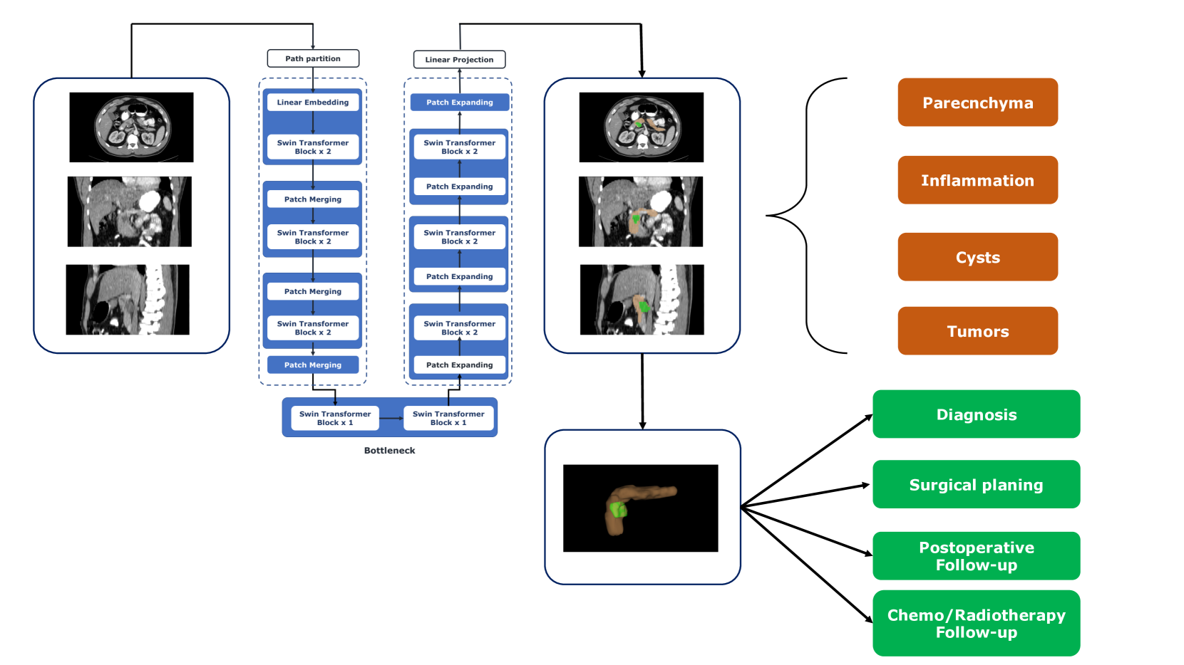
Pancreas segmentation has been traditionally challenging due to its small size in computed tomography abdominal volumes, high variability of shape and positions among patients, and blurred boundaries due to low contrast between the pancreas and surrounding organs. Many deep learning models for pancreas segmentation have been proposed in the past few years. We present a thorough systematic review based on the Preferred Reporting Items for Systematic Reviews and Meta-analyses (PRISMA) statement. The literature search was conducted on PubMed, Web of Science, Scopus, and IEEE Xplore on original studies published in peer-reviewed journals from 2013 to 2023. Overall, 130 studies were retrieved. We initially provided an overview of the technical background of the most common network architectures and publicly available datasets. Then, the analysis of the studies combining visual presentation in tabular form and text description was reported. The tables grouped the studies specifying the application, dataset size, design (model architecture, learning strategy, and loss function), results, and main contributions. We first analyzed the studies focusing on parenchyma segmentation using coarse-to-fine approaches, multi-organ segmentation, semi-supervised learning, and unsupervised learning, followed by those studies on generalization to other datasets and those concerning the design of new loss functions. Then, we analyzed the studies on segmentation of tumors, cysts, and inflammation reporting multi-stage methods, semi-supervised learning, generalization to other datasets, and design of new loss functions. Finally, we provided a critical discussion on the subject based on the published evidence underlining current issues that need to be addressed before clinical translation.
Read more7/24/2024
🤿
0
Large-Scale Multi-Center CT and MRI Segmentation of Pancreas with Deep Learning
Zheyuan Zhang, Elif Keles, Gorkem Durak, Yavuz Taktak, Onkar Susladkar, Vandan Gorade, Debesh Jha, Asli C. Ormeci, Alpay Medetalibeyoglu, Lanhong Yao, Bin Wang, Ilkin Sevgi Isler, Linkai Peng, Hongyi Pan, Camila Lopes Vendrami, Amir Bourhani, Yury Velichko, Boqing Gong, Concetto Spampinato, Ayis Pyrros, Pallavi Tiwari, Derk C. F. Klatte, Megan Engels, Sanne Hoogenboom, Candice W. Bolan, Emil Agarunov, Nassier Harfouch, Chenchan Huang, Marco J. Bruno, Ivo Schoots, Rajesh N. Keswani, Frank H. Miller, Tamas Gonda, Cemal Yazici, Temel Tirkes, Baris Turkbey, Michael B. Wallace, Ulas Bagci
Automated volumetric segmentation of the pancreas on cross-sectional imaging is needed for diagnosis and follow-up of pancreatic diseases. While CT-based pancreatic segmentation is more established, MRI-based segmentation methods are understudied, largely due to a lack of publicly available datasets, benchmarking research efforts, and domain-specific deep learning methods. In this retrospective study, we collected a large dataset (767 scans from 499 participants) of T1-weighted (T1W) and T2-weighted (T2W) abdominal MRI series from five centers between March 2004 and November 2022. We also collected CT scans of 1,350 patients from publicly available sources for benchmarking purposes. We developed a new pancreas segmentation method, called PanSegNet, combining the strengths of nnUNet and a Transformer network with a new linear attention module enabling volumetric computation. We tested PanSegNet's accuracy in cross-modality (a total of 2,117 scans) and cross-center settings with Dice and Hausdorff distance (HD95) evaluation metrics. We used Cohen's kappa statistics for intra and inter-rater agreement evaluation and paired t-tests for volume and Dice comparisons, respectively. For segmentation accuracy, we achieved Dice coefficients of 88.3% (std: 7.2%, at case level) with CT, 85.0% (std: 7.9%) with T1W MRI, and 86.3% (std: 6.4%) with T2W MRI. There was a high correlation for pancreas volume prediction with R^2 of 0.91, 0.84, and 0.85 for CT, T1W, and T2W, respectively. We found moderate inter-observer (0.624 and 0.638 for T1W and T2W MRI, respectively) and high intra-observer agreement scores. All MRI data is made available at https://osf.io/kysnj/. Our source code is available at https://github.com/NUBagciLab/PaNSegNet.
Read more5/28/2024

0
Automatic Organ and Pan-cancer Segmentation in Abdomen CT: the FLARE 2023 Challenge
Jun Ma, Yao Zhang, Song Gu, Cheng Ge, Ershuai Wang, Qin Zhou, Ziyan Huang, Pengju Lyu, Jian He, Bo Wang
Organ and cancer segmentation in abdomen Computed Tomography (CT) scans is the prerequisite for precise cancer diagnosis and treatment. Most existing benchmarks and algorithms are tailored to specific cancer types, limiting their ability to provide comprehensive cancer analysis. This work presents the first international competition on abdominal organ and pan-cancer segmentation by providing a large-scale and diverse dataset, including 4650 CT scans with various cancer types from over 40 medical centers. The winning team established a new state-of-the-art with a deep learning-based cascaded framework, achieving average Dice Similarity Coefficient scores of 92.3% for organs and 64.9% for lesions on the hidden multi-national testing set. The dataset and code of top teams are publicly available, offering a benchmark platform to drive further innovations https://codalab.lisn.upsaclay.fr/competitions/12239.
Read more8/23/2024

0
Optimizing Synthetic Data for Enhanced Pancreatic Tumor Segmentation
Linkai Peng, Zheyuan Zhang, Gorkem Durak, Frank H. Miller, Alpay Medetalibeyoglu, Michael B. Wallace, Ulas Bagci
Pancreatic cancer remains one of the leading causes of cancer-related mortality worldwide. Precise segmentation of pancreatic tumors from medical images is a bottleneck for effective clinical decision-making. However, achieving a high accuracy is often limited by the small size and availability of real patient data for training deep learning models. Recent approaches have employed synthetic data generation to augment training datasets. While promising, these methods may not yet meet the performance benchmarks required for real-world clinical use. This study critically evaluates the limitations of existing generative-AI based frameworks for pancreatic tumor segmentation. We conduct a series of experiments to investigate the impact of synthetic textit{tumor size} and textit{boundary definition} precision on model performance. Our findings demonstrate that: (1) strategically selecting a combination of synthetic tumor sizes is crucial for optimal segmentation outcomes, and (2) generating synthetic tumors with precise boundaries significantly improves model accuracy. These insights highlight the importance of utilizing refined synthetic data augmentation for enhancing the clinical utility of segmentation models in pancreatic cancer decision making including diagnosis, prognosis, and treatment plans. Our code will be available at https://github.com/lkpengcs/SynTumorAnalyzer.
Read more7/30/2024