Boundary Constraint-free Biomechanical Model-Based Surface Matching for Intraoperative Liver Deformation Correction

0
Sign in to get full access
Overview
- The paper presents a biomechanical model-based approach for correcting intraoperative liver deformation during image-guided surgery.
- It proposes a boundary constraint-free surface matching technique that aligns pre-operative and intraoperative liver surfaces without the need for boundary constraints.
- The method aims to improve the accuracy of non-rigid liver registration and deformation correction for enhanced surgical guidance.
Plain English Explanation
When performing surgery on the liver, the shape and position of the liver can change significantly from the pre-operative (before surgery) images to the actual surgery. This can make it challenging to accurately guide the surgery based on the pre-operative images.
The researchers in this paper developed a new technique to better align the pre-operative liver image with the actual liver during surgery. Their approach uses a biomechanical model of the liver to simulate and predict how the liver will deform and change shape during the surgery. This biomechanical model is then used to match the pre-operative liver image to the actual liver surface observed during surgery, without needing to constrain the liver boundaries.
By avoiding the need for boundary constraints, this method can more accurately capture the complex deformations the liver undergoes during the procedure. This improved alignment between the pre-operative image and the actual liver shape can then be used to better guide the surgeon and ensure the surgery is performed accurately.
Technical Explanation
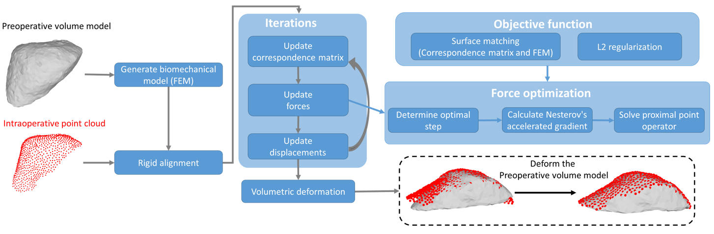
The paper proposes a biomechanical model-based surface matching approach for correcting intraoperative liver deformation. It uses a finite element liver model to simulate the deformation of the pre-operative liver surface and match it to the intraoperative liver surface observed during surgery.
The key aspects of the method are:
-
Biomechanical Liver Modeling: The researchers develop a patient-specific finite element model of the liver using the pre-operative CT/MRI data. This model captures the mechanical properties and boundary conditions of the liver.
-
Boundary Constraint-free Surface Matching: Instead of relying on boundary constraints, the method uses an optimization-based approach to deform the pre-operative liver surface to align with the intraoperative surface observations. This allows for more flexibility in capturing complex liver deformations.
-
Deformation Correction: Once the pre-operative and intraoperative liver surfaces are aligned, the method can then use the biomechanical model to compute the full 3D deformation field, enabling accurate correction of the pre-operative image for enhanced surgical guidance.
Critical Analysis
The proposed method is a valuable contribution to the field of image-guided liver surgery, as it addresses the challenge of accurately capturing complex liver deformations during the procedure. By avoiding the need for boundary constraints, the approach can better handle the unpredictable nature of intraoperative liver deformation.
However, the paper does not extensively discuss the computational complexity and runtime of the optimization-based surface matching algorithm. This could be an important practical consideration, as real-time performance is crucial for effective surgical guidance.
Additionally, the evaluation is limited to a small patient cohort, and further validation on a larger and more diverse dataset would be beneficial to assess the generalizability of the method.
Conclusion
This paper presents a promising biomechanical model-based approach for correcting intraoperative liver deformation during image-guided surgery. By using a boundary constraint-free surface matching technique, the method can better capture the complex deformations of the liver, leading to improved registration accuracy and enhanced surgical guidance.
The proposed approach represents an important step forward in addressing the challenges of non-rigid liver registration, and its potential impact could be significant in improving the outcomes of liver surgery procedures.
This summary was produced with help from an AI and may contain inaccuracies - check out the links to read the original source documents!
Related Papers
0
Boundary Constraint-free Biomechanical Model-Based Surface Matching for Intraoperative Liver Deformation Correction
Zixin Yang, Richard Simon, Kelly Merrell, Cristian. A. Linte
In image-guided liver surgery, 3D-3D non-rigid registration methods play a crucial role in estimating the mapping between the preoperative model and the intraoperative surface represented as point clouds, addressing the challenge of tissue deformation. Typically, these methods incorporate a biomechanical model, represented as a finite element model (FEM), used to regularize a surface matching term. This paper introduces a novel 3D-3D non-rigid registration method. In contrast to the preceding techniques, our method uniquely incorporates the FEM within the surface matching term itself, ensuring that the estimated deformation maintains geometric consistency throughout the registration process. Additionally, we eliminate the need to determine zero-boundary conditions and applied force locations in the FEM. We achieve this by integrating soft springs into the stiffness matrix and allowing forces to be distributed across the entire liver surface. To further improve robustness, we introduce a regularization technique focused on the gradient of the force magnitudes. This regularization imposes spatial smoothness and helps prevent the overfitting of irregular noise in intraoperative data. Optimization is achieved through an accelerated proximal gradient algorithm, further enhanced by our proposed method for determining the optimal step size. Our method is evaluated and compared to both a learning-based method and a traditional method that features FEM regularization using data collected on our custom-developed phantom, as well as two publicly available datasets. Our method consistently outperforms or is comparable to the baseline techniques. Both the code and dataset will be made publicly available.
Read more9/10/2024
🖼️
0
Diffeomorphic Transformer-based Abdomen MRI-CT Deformable Image Registration
Yang Lei, Luke A. Matkovic, Justin Roper, Tonghe Wang, Jun Zhou, Beth Ghavidel, Mark McDonald, Pretesh Patel, Xiaofeng Yang
This paper aims to create a deep learning framework that can estimate the deformation vector field (DVF) for directly registering abdominal MRI-CT images. The proposed method assumed a diffeomorphic deformation. By using topology-preserved deformation features extracted from the probabilistic diffeomorphic registration model, abdominal motion can be accurately obtained and utilized for DVF estimation. The model integrated Swin transformers, which have demonstrated superior performance in motion tracking, into the convolutional neural network (CNN) for deformation feature extraction. The model was optimized using a cross-modality image similarity loss and a surface matching loss. To compute the image loss, a modality-independent neighborhood descriptor (MIND) was used between the deformed MRI and CT images. The surface matching loss was determined by measuring the distance between the warped coordinates of the surfaces of contoured structures on the MRI and CT images. The deformed MRI image was assessed against the CT image using the target registration error (TRE), Dice similarity coefficient (DSC), and mean surface distance (MSD) between the deformed contours of the MRI image and manual contours of the CT image. When compared to only rigid registration, DIR with the proposed method resulted in an increase of the mean DSC values of the liver and portal vein from 0.850 and 0.628 to 0.903 and 0.763, a decrease of the mean MSD of the liver from 7.216 mm to 3.232 mm, and a decrease of the TRE from 26.238 mm to 8.492 mm. The proposed deformable image registration method based on a diffeomorphic transformer provides an effective and efficient way to generate an accurate DVF from an MRI-CT image pair of the abdomen. It could be utilized in the current treatment planning workflow for liver radiotherapy.
Read more5/7/2024

0
Biomechanics-informed Non-rigid Medical Image Registration and its Inverse Material Property Estimation with Linear and Nonlinear Elasticity
Zhe Min, Zachary M. C. Baum, Shaheer U. Saeed, Mark Emberton, Dean C. Barratt, Zeike A. Taylor, Yipeng Hu
This paper investigates both biomechanical-constrained non-rigid medical image registrations and accurate identifications of material properties for soft tissues, using physics-informed neural networks (PINNs). The complex nonlinear elasticity theory is leveraged to formally establish the partial differential equations (PDEs) representing physics laws of biomechanical constraints that need to be satisfied, with which registration and identification tasks are treated as forward (i.e., data-driven solutions of PDEs) and inverse (i.e., parameter estimation) problems under PINNs respectively. Two net configurations (i.e., Cfg1 and Cfg2) have also been compared for both linear and nonlinear physics model. Two sets of experiments have been conducted, using pairs of undeformed and deformed MR images from clinical cases of prostate cancer biopsy. Our contributions are summarised as follows. 1) We developed a learning-based biomechanical-constrained non-rigid registration algorithm using PINNs, where linear elasticity is generalised to the nonlinear version. 2) We demonstrated extensively that nonlinear elasticity shows no statistical significance against linear models in computing point-wise displacement vectors but their respective benefits may depend on specific patients, with finite-element (FE) computed ground-truth. 3) We formulated and solved the inverse parameter estimation problem, under the joint optimisation scheme of registration and parameter identification using PINNs, whose solutions can be accurately found by locating saddle points.
Read more7/10/2024

0
Data-Driven Tissue- and Subject-Specific Elastic Regularization for Medical Image Registration
Anna Reithmeir, Lina Felsner, Rickmer Braren, Julia A. Schnabel, Veronika A. Zimmer
Physics-inspired regularization is desired for intra-patient image registration since it can effectively capture the biomechanical characteristics of anatomical structures. However, a major challenge lies in the reliance on physical parameters: Parameter estimations vary widely across the literature, and the physical properties themselves are inherently subject-specific. In this work, we introduce a novel data-driven method that leverages hypernetworks to learn the tissue-dependent elasticity parameters of an elastic regularizer. Notably, our approach facilitates the estimation of patient-specific parameters without the need to retrain the network. We evaluate our method on three publicly available 2D and 3D lung CT and cardiac MR datasets. We find that with our proposed subject-specific tissue-dependent regularization, a higher registration quality is achieved across all datasets compared to using a global regularizer. The code is available at https://github.com/compai-lab/2024-miccai-reithmeir.
Read more7/8/2024