Diffeomorphic Transformer-based Abdomen MRI-CT Deformable Image Registration

0
🖼️
Sign in to get full access
Overview
- The paper proposes a deep learning framework to estimate the deformation vector field (DVF) for direct registration of abdominal MRI-CT images.
- The method assumes a diffeomorphic deformation, using topology-preserved deformation features extracted from a probabilistic diffeomorphic registration model to accurately obtain abdominal motion for DVF estimation.
- The model integrates Swin transformers, known for their superior performance in motion tracking, into a convolutional neural network (CNN) for deformation feature extraction.
- The model is optimized using a cross-modality image similarity loss and a surface matching loss, leveraging a modality-independent neighborhood descriptor (MIND) and measuring the distance between warped and manual contours.
- Evaluation shows the proposed method outperforms rigid registration, with improvements in Dice similarity coefficient (DSC), mean surface distance (MSD), and target registration error (TRE).
Plain English Explanation
The researchers developed a machine learning system that can accurately align abdominal MRI and CT images. This is important for medical applications like radiation therapy planning, where having precise alignment between the two image modalities is crucial.
The key innovation is the use of a Transformer-based model to extract features that describe the deformation, or warping, between the MRI and CT images. This allows the system to estimate a detailed "deformation vector field" that maps how each part of the MRI image needs to be transformed to match the CT image.
To train the model, the researchers used a combination of two loss functions. The first measures how well the deformed MRI image matches the CT image, using a special technique called MIND that can compare images from different modalities. The second loss function measures how well the deformed surfaces of anatomical structures in the MRI match the manual contours in the CT image.
Compared to a simpler rigid registration approach, the proposed deformable image registration method was able to achieve significantly better alignment, as shown by improvements in various evaluation metrics like Dice similarity and target registration error.
Technical Explanation
The paper proposes a diffeomorphic transformer-based deformable image registration method for aligning abdominal MRI and CT images. The key technical components include:
- Diffeomorphic Deformation: The method assumes a diffeomorphic (smooth and invertible) deformation model, which is well-suited for capturing the complex nonlinear motion of abdominal organs.
- Deformation Feature Extraction: The researchers integrate Swin transformers, known for their performance in motion tracking, into a CNN to extract informative deformation features from the images.
- Cross-Modality Similarity Loss: To compare the deformed MRI to the CT image, the method uses a modality-independent neighborhood descriptor (MIND) to compute the image similarity loss in a way that is robust to the differences between MRI and CT.
- Surface Matching Loss: Additionally, the model optimizes a surface matching loss by measuring the distance between the deformed contours of anatomical structures in the MRI and the manual contours in the CT.
The proposed diffeomorphic registration approach outperformed a standard rigid registration method, demonstrating improved Dice similarity, mean surface distance, and target registration error on the abdominal MRI-CT alignment task.
Critical Analysis
The paper presents a well-designed and thorough evaluation of the proposed method, including comparisons to a rigid registration baseline and detailed analyses of the registration accuracy for different anatomical structures. However, a few potential limitations and areas for further research are worth noting:
- The method was only evaluated on a dataset of abdominal MRI-CT image pairs, so its generalization to other body regions or modality combinations is unclear. Further testing on a broader range of medical imaging data would help establish the method's broader applicability.
- The paper does not provide much insight into the failure cases or specific situations where the deformable registration method may struggle. Understanding the limitations and robustness of the approach in challenging scenarios would be valuable.
- While the transformer-based deformation feature extraction is a key innovation, the paper does not provide a detailed ablation study to quantify the specific contributions of this component compared to other design choices.
Overall, the proposed deep learning framework for MRI-CT deformable image registration appears to be a promising approach, but further research and testing would help solidify its strengths and limitations.
Conclusion
This paper presents an effective deep learning framework for estimating the deformation vector field (DVF) to directly register abdominal MRI and CT images. By leveraging a diffeomorphic deformation model and integrating Swin transformers into a CNN, the method is able to accurately capture the complex nonlinear motion of abdominal organs.
The proposed approach outperforms rigid registration, demonstrating significant improvements in key evaluation metrics like Dice similarity, mean surface distance, and target registration error. This could have important implications for medical applications like radiation therapy planning, where precise alignment between MRI and CT images is critical.
While further research is needed to fully assess the method's generalization and robustness, this work represents an important step forward in developing effective deep learning solutions for multimodal deformable image registration in the medical domain.
This summary was produced with help from an AI and may contain inaccuracies - check out the links to read the original source documents!
Related Papers
🖼️
0
Diffeomorphic Transformer-based Abdomen MRI-CT Deformable Image Registration
Yang Lei, Luke A. Matkovic, Justin Roper, Tonghe Wang, Jun Zhou, Beth Ghavidel, Mark McDonald, Pretesh Patel, Xiaofeng Yang
This paper aims to create a deep learning framework that can estimate the deformation vector field (DVF) for directly registering abdominal MRI-CT images. The proposed method assumed a diffeomorphic deformation. By using topology-preserved deformation features extracted from the probabilistic diffeomorphic registration model, abdominal motion can be accurately obtained and utilized for DVF estimation. The model integrated Swin transformers, which have demonstrated superior performance in motion tracking, into the convolutional neural network (CNN) for deformation feature extraction. The model was optimized using a cross-modality image similarity loss and a surface matching loss. To compute the image loss, a modality-independent neighborhood descriptor (MIND) was used between the deformed MRI and CT images. The surface matching loss was determined by measuring the distance between the warped coordinates of the surfaces of contoured structures on the MRI and CT images. The deformed MRI image was assessed against the CT image using the target registration error (TRE), Dice similarity coefficient (DSC), and mean surface distance (MSD) between the deformed contours of the MRI image and manual contours of the CT image. When compared to only rigid registration, DIR with the proposed method resulted in an increase of the mean DSC values of the liver and portal vein from 0.850 and 0.628 to 0.903 and 0.763, a decrease of the mean MSD of the liver from 7.216 mm to 3.232 mm, and a decrease of the TRE from 26.238 mm to 8.492 mm. The proposed deformable image registration method based on a diffeomorphic transformer provides an effective and efficient way to generate an accurate DVF from an MRI-CT image pair of the abdomen. It could be utilized in the current treatment planning workflow for liver radiotherapy.
Read more5/7/2024

0
Gaussian Representation for Deformable Image Registration
Jihe Li, Fabian Zhang, Xia Li, Tianhao Zhang, Ye Zhang, Joachim Buhmann
Deformable image registration (DIR) is a fundamental task in radiotherapy, with existing methods often struggling to balance computational efficiency, registration accuracy, and speed effectively. We introduce a novel DIR approach employing parametric 3D Gaussian control points achieving a better tradeoff. It provides an explicit and flexible representation for spatial deformation fields between 3D volumetric medical images, producing a displacement vector field (DVF) across all volumetric positions. The movement of individual voxels is derived using linear blend skinning (LBS) through localized interpolation of transformations associated with neighboring Gaussians. This interpolation strategy not only simplifies the determination of voxel motions but also acts as an effective regularization technique. Our approach incorporates a unified optimization process through backpropagation, enabling iterative learning of both the parameters of the 3D Gaussians and their transformations. Additionally, the density of Gaussians is adjusted adaptively during the learning phase to accommodate varying degrees of motion complexity. We validated our approach on the 4D-CT lung DIR-Lab and cardiac ACDC datasets, achieving an average target registration error (TRE) of 1.06 mm within a much-improved processing time of 2.43 seconds for the DIR-Lab dataset over existing methods, demonstrating significant advancements in both accuracy and efficiency.
Read more6/6/2024
🧠
0
Efficient Post-processing of Diffusion Tensor Cardiac Magnetic Imaging Using Texture-conserving Deformable Registration
Fanwen Wang, Pedro F. Ferreira, Yinzhe Wu, Camila Munoz, Ke Wen, Yaqing Luo, Jiahao Huang, Dudley J. Pennell, Andrew D. Scott, Sonia Nielles-Vallespin, Guang Yang
Diffusion tensor cardiac magnetic resonance (DT-CMR) is a method capable of providing non-invasive measurements of myocardial microstructure. Image registration is essential to correct image shifts due to intra and inter breath-hold motion and imperfect cardiac triggering. Registration is challenging in DT-CMR due to the low signal-to-noise and various contrasts induced by the diffusion encoding in the myocardium and surrounding organs. Traditional deformable registration corrects through-plane motion but at the risk of destroying the texture information while rigid registration inefficiently discards frames with local deformation. In this study, we explored the possibility of deep learning-based deformable registration on DT-CMR. Based on the noise suppression using low-rank features and diffusion encoding suppression using variational auto encoder-decoder, a B-spline based registration network extracted the displacement fields and maintained the texture features of DT-CMR. In this way, our method improved the efficiency of frame utilization, manual cropping, and computational speed.
Read more5/17/2024
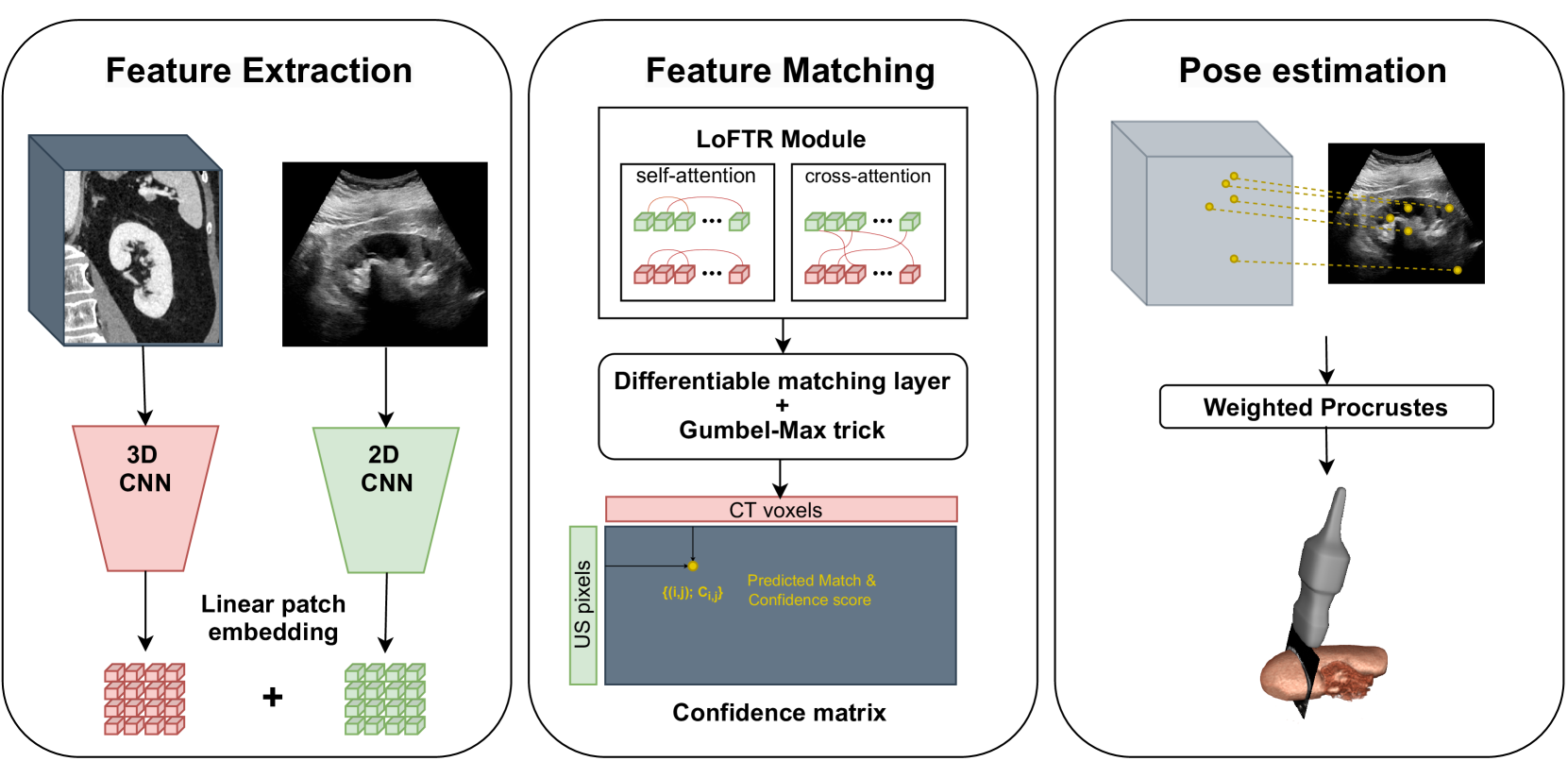
0
Transformer-Based Local Feature Matching for Multimodal Image Registration
Remi Delaunay, Ruisi Zhang, Filipe C. Pedrosa, Navid Feizi, Dianne Sacco, Rajni Patel, Jayender Jagadeesan
Ultrasound imaging is a cost-effective and radiation-free modality for visualizing anatomical structures in real-time, making it ideal for guiding surgical interventions. However, its limited field-of-view, speckle noise, and imaging artifacts make it difficult to interpret the images for inexperienced users. In this paper, we propose a new 2D ultrasound to 3D CT registration method to improve surgical guidance during ultrasound-guided interventions. Our approach adopts a dense feature matching method called LoFTR to our multimodal registration problem. We learn to predict dense coarse-to-fine correspondences using a Transformer-based architecture to estimate a robust rigid transformation between a 2D ultrasound frame and a CT scan. Additionally, a fully differentiable pose estimation method is introduced, optimizing LoFTR on pose estimation error during training. Experiments conducted on a multimodal dataset of ex vivo porcine kidneys demonstrate the method's promising results for intraoperative, trackerless ultrasound pose estimation. By mapping 2D ultrasound frames into the 3D CT volume space, the method provides intraoperative guidance, potentially improving surgical workflows and image interpretation.
Read more4/26/2024