Brain Tumor Classification From MRI Images Using Machine Learning

0
🏷️
Sign in to get full access
Overview
- Brain tumors are a serious medical condition that can disrupt normal brain function
- The average 5-year survival rate for malignant brain tumors is only 35.6%
- Early detection is crucial for effective treatment planning
- Advances in medical imaging technology, like MRI scans, enable better diagnosis
- Machine learning and deep learning algorithms show promise for automated brain tumor detection and classification
Plain English Explanation
Brain tumors are abnormal growths that develop in the brain and can be life-threatening. They can interfere with the normal functioning of the brain, which is a serious problem. Unfortunately, the average 5-year survival rate for malignant (cancerous) brain tumors is only 35.6%.
To improve patient outcomes, it's important to detect brain tumors as early as possible so that doctors can develop an effective treatment plan. Advancements in medical imaging technology, such as magnetic resonance imaging (MRI) scans, have made it easier to get detailed images of the brain. Enhancing brain tumor segmentation across diverse populations and optimized ensemble deep learning model for brain tumor are two examples of research exploring how to use these brain scans more effectively.
Researchers have started to explore using machine learning and deep learning algorithms to automatically analyze brain scans and detect the presence of tumors. Deep learning-based brain image segmentation and exploration of multi-scale image fusion systems are two papers that investigate these techniques. The goal is to create predictive systems that can help doctors identify and classify brain tumors more accurately and efficiently.
Technical Explanation
The paper discusses developing a predictive system for brain tumor detection using machine learning techniques, specifically ensemble methods. Ensemble learning combines multiple individual machine learning models to improve the overall performance and robustness of the system.
The researchers propose an approach that leverages different machine learning algorithms, including decision trees, random forests, and support vector machines, to create an ensemble model for brain tumor classification. They use magnetic resonance imaging (MRI) scans as the input data and extract relevant features from the images to train the ensemble model.
The brain MRI detection by semantic segmentation models paper explores a similar approach, using deep learning-based semantic segmentation to identify and localize brain tumors in MRI scans.
The key insight from this research is that ensemble methods can outperform individual machine learning models in the task of brain tumor detection and classification. By combining the strengths of different algorithms, the ensemble approach can provide more accurate and reliable predictions, which is crucial for early diagnosis and effective treatment planning.
Critical Analysis
The research presented in the paper provides a promising approach for brain tumor detection using ensemble machine learning techniques. However, it's important to consider some potential limitations and areas for further exploration.
One limitation is the reliance on MRI scans as the sole input data. While MRI is a powerful imaging modality, incorporating additional data sources, such as genomic information or clinical data, could potentially improve the model's performance and provide a more comprehensive understanding of brain tumor characteristics.
Additionally, the paper does not address the challenge of tumor localization and classification. Accurately identifying the location and type of brain tumor within the scan is a crucial step for treatment planning. Exploration of multi-scale image fusion systems and brain MRI detection by semantic segmentation models demonstrate techniques that address these challenges.
Further research could also investigate the performance of the ensemble model across diverse patient populations and explore ways to enhance its generalization capabilities. Ensuring the model's robustness and fairness is essential for real-world clinical applications.
Conclusion
This research paper presents a promising approach for brain tumor detection using ensemble machine learning techniques. By leveraging the strengths of different algorithms, the ensemble model can provide more accurate and reliable predictions, which is critical for early diagnosis and effective treatment planning.
While the research demonstrates the potential of this approach, there are opportunities for further exploration, such as incorporating additional data sources, addressing tumor localization and classification, and ensuring the model's robustness and fairness across diverse patient populations. Ongoing advancements in medical imaging and machine learning will continue to drive progress in this important field, ultimately leading to improved patient outcomes.
This summary was produced with help from an AI and may contain inaccuracies - check out the links to read the original source documents!
Related Papers
🏷️
0
Brain Tumor Classification From MRI Images Using Machine Learning
Vidhyapriya Ranganathan, Celshiya Udaiyar, Jaisree Jayanth, Meghaa P V, Srija B, Uthra S
Brain tumor is a life-threatening problem and hampers the normal functioning of the human body. The average five-year relative survival rate for malignant brain tumors is 35.6 percent. For proper diagnosis and efficient treatment planning, it is necessary to detect the brain tumor in early stages. Due to advancement in medical imaging technology, the brain images are taken in different modalities. The ability to extract relevant characteristics from magnetic resonance imaging (MRI) scans is a crucial step for brain tumor classifiers. Several studies have proposed various strategies to extract relevant features from different modalities of MRI to predict the growth of abnormal tumors. Most techniques used conventional methods of image processing for feature extraction and machine learning for classification. More recently, the use of deep learning algorithms in medical imaging has resulted in significant improvements in the classification and diagnosis of brain tumors. Since tumors are located at different regions of the brain, localizing the tumor and classifying it to a particular category is a challenging task. The objective of this project is to develop a predictive system for brain tumor detection using machine learning(ensembling).
Read more7/16/2024

0
Deep Learning in Medical Image Classification from MRI-based Brain Tumor Images
Xiaoyi Liu, Zhuoyue Wang
Brain tumors are among the deadliest diseases in the world. Magnetic Resonance Imaging (MRI) is one of the most effective ways to detect brain tumors. Accurate detection of brain tumors based on MRI scans is critical, as it can potentially save many lives and facilitate better decision-making at the early stages of the disease. Within our paper, four different types of MRI-based images have been collected from the database: glioma tumor, no tumor, pituitary tumor, and meningioma tumor. Our study focuses on making predictions for brain tumor classification. Five models, including four pre-trained models (MobileNet, EfficientNet-B0, ResNet-18, and VGG16) and one new model, MobileNet-BT, have been proposed for this study.
Read more8/2/2024
0
On Enhancing Brain Tumor Segmentation Across Diverse Populations with Convolutional Neural Networks
Fadillah Maani, Anees Ur Rehman Hashmi, Numan Saeed, Mohammad Yaqub
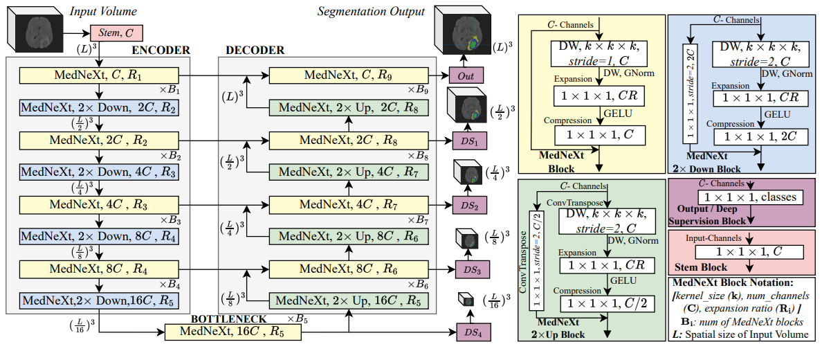
Brain tumor segmentation is a fundamental step in assessing a patient's cancer progression. However, manual segmentation demands significant expert time to identify tumors in 3D multimodal brain MRI scans accurately. This reliance on manual segmentation makes the process prone to intra- and inter-observer variability. This work proposes a brain tumor segmentation method as part of the BraTS-GoAT challenge. The task is to segment tumors in brain MRI scans automatically from various populations, such as adults, pediatrics, and underserved sub-Saharan Africa. We employ a recent CNN architecture for medical image segmentation, namely MedNeXt, as our baseline, and we implement extensive model ensembling and postprocessing for inference. Our experiments show that our method performs well on the unseen validation set with an average DSC of 85.54% and HD95 of 27.88. The code is available on https://github.com/BioMedIA-MBZUAI/BraTS2024_BioMedIAMBZ.
Read more5/7/2024
🤿
0
An Optimized Ensemble Deep Learning Model For Brain Tumor Classification
Md. Alamin Talukder, Md. Manowarul Islam, Md Ashraf Uddin
Brain tumors present a grave risk to human life, demanding precise and timely diagnosis for effective treatment. Inaccurate identification of brain tumors can significantly diminish life expectancy, underscoring the critical need for precise diagnostic methods. Manual identification of brain tumors within vast Magnetic Resonance Imaging (MRI) image datasets is arduous and time-consuming. Thus, the development of a reliable deep learning (DL) model is essential to enhance diagnostic accuracy and ultimately save lives. This study introduces an innovative optimization-based deep ensemble approach employing transfer learning (TL) to efficiently classify brain tumors. Our methodology includes meticulous preprocessing, reconstruction of TL architectures, fine-tuning, and ensemble DL models utilizing weighted optimization techniques such as Genetic Algorithm-based Weight Optimization (GAWO) and Grid Search-based Weight Optimization (GSWO). Experimentation is conducted on the Figshare Contrast-Enhanced MRI (CE-MRI) brain tumor dataset, comprising 3064 images. Our approach achieves notable accuracy scores, with Xception, ResNet50V2, ResNet152V2, InceptionResNetV2, GAWO, and GSWO attaining 99.42%, 98.37%, 98.22%, 98.26%, 99.71%, and 99.76% accuracy, respectively. Notably, GSWO demonstrates superior accuracy, averaging 99.76% accuracy across five folds on the Figshare CE-MRI brain tumor dataset. The comparative analysis highlights the significant performance enhancement of our proposed model over existing counterparts. In conclusion, our optimized deep ensemble model exhibits exceptional accuracy in swiftly classifying brain tumors. Furthermore, it has the potential to assist neurologists and clinicians in making accurate and immediate diagnostic decisions.
Read more5/7/2024