CAMEL2: Enhancing weakly supervised learning for histopathology images by incorporating the significance ratio

0

Sign in to get full access
Overview
- This paper introduces CAMEL2, a method for enhancing weakly supervised learning for histopathology image analysis.
- The key innovation is the incorporation of a "significance ratio" to better utilize the limited labeled data available in weakly supervised settings.
- CAMEL2 builds on previous work like CAMEL, Self-Contrastive, and Morphology-Enhanced CAM.
Plain English Explanation
Histopathology, the microscopic study of diseased tissues, is a crucial tool for cancer diagnosis. However, labeling and annotating these medical images can be time-consuming and expensive. To address this, researchers have developed "weakly supervised" learning techniques that can train models using only partial or imprecise labels.
The CAMEL2 method builds on this idea by incorporating a novel "significance ratio" metric. This ratio helps the model focus on the most informative regions of the histopathology images, even when the labels are incomplete. By emphasizing the most relevant visual features, CAMEL2 can learn more effectively from limited labeled data.
This is similar to how humans might learn - we often focus our attention on the most important details when trying to understand a new concept with limited information. CAMEL2 mimics this selective attention to extract the most valuable insights from weakly labeled medical images.
Technical Explanation
The CAMEL2 method consists of several key components:
-
Weakly Supervised Learning: CAMEL2 utilizes a weakly supervised setup, where the model is trained on images with imprecise or incomplete labels, rather than detailed annotations of every relevant region.
-
Significance Ratio Calculation: A key innovation in CAMEL2 is the incorporation of a "significance ratio" for each image region. This ratio measures how informative a given region is for the target classification task, relative to the rest of the image. Regions with a higher significance ratio are weighted more heavily during training.
-
Weakly Supervised Attention Mechanism: CAMEL2 employs a weakly supervised attention mechanism that focuses the model's attention on the most significant regions of the histopathology images, as determined by the calculated significance ratios.
-
Iterative Refinement: The CAMEL2 training process involves iteratively refining the significance ratios and attention mechanism, allowing the model to progressively learn from the most informative image regions.
By leveraging the significance ratio to guide the weakly supervised attention mechanism, CAMEL2 is able to extract more valuable information from limited labeled data compared to previous approaches. This enhances the model's performance on histopathology image classification tasks.
Critical Analysis
The CAMEL2 paper makes a valuable contribution to the field of weakly supervised learning for medical image analysis. The significance ratio concept is a clever way to focus the model's attention on the most relevant visual features, even when the training labels are incomplete or imprecise.
However, the authors acknowledge that CAMEL2 is still limited by the inherent challenges of weakly supervised learning, such as the potential for the model to latch onto spurious correlations in the data. Additionally, the iterative refinement process, while effective, may increase the computational complexity and training time of the method.
Further research could explore ways to make the significance ratio calculation more robust, or to integrate CAMEL2 with other techniques like self-supervised learning or region-of-interest detection to address these limitations.
Conclusion
The CAMEL2 method represents a promising advancement in weakly supervised learning for histopathology image analysis. By incorporating a significance ratio to guide the attention mechanism, CAMEL2 can learn more effectively from limited labeled data, with potential applications in cancer detection and prognostic modeling.
As medical imaging datasets continue to grow, techniques like CAMEL2 will become increasingly important for leveraging these resources to advance our understanding and treatment of disease.
This summary was produced with help from an AI and may contain inaccuracies - check out the links to read the original source documents!
Related Papers
0
CAMEL2: Enhancing weakly supervised learning for histopathology images by incorporating the significance ratio
Gang Xu, Shuhao Wang, Lingyu Zhao, Xiao Chen, Tongwei Wang, Lang Wang, Zhenwei Luo, Dahan Wang, Zewen Zhang, Aijun Liu, Wei Ba, Zhigang Song, Huaiyin Shi, Dingrong Zhong, Jianpeng Ma
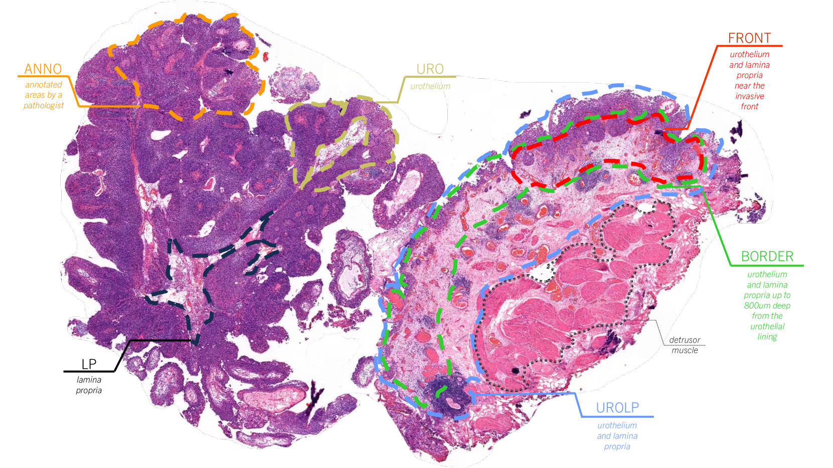
Histopathology image analysis plays a crucial role in cancer diagnosis. However, training a clinically applicable segmentation algorithm requires pathologists to engage in labour-intensive labelling. In contrast, weakly supervised learning methods, which only require coarse-grained labels at the image level, can significantly reduce the labeling efforts. Unfortunately, while these methods perform reasonably well in slide-level prediction, their ability to locate cancerous regions, which is essential for many clinical applications, remains unsatisfactory. Previously, we proposed CAMEL, which achieves comparable results to those of fully supervised baselines in pixel-level segmentation. However, CAMEL requires 1,280x1,280 image-level binary annotations for positive WSIs. Here, we present CAMEL2, by introducing a threshold of the cancerous ratio for positive bags, it allows us to better utilize the information, consequently enabling us to scale up the image-level setting from 1,280x1,280 to 5,120x5,120 while maintaining the accuracy. Our results with various datasets, demonstrate that CAMEL2, with the help of 5,120x5,120 image-level binary annotations, which are easy to annotate, achieves comparable performance to that of a fully supervised baseline in both instance- and slide-level classifications.
Read more5/28/2024
🔎
0
Boosting Medical Image-based Cancer Detection via Text-guided Supervision from Reports
Guangyu Guo, Jiawen Yao, Yingda Xia, Tony C. W. Mok, Zhilin Zheng, Junwei Han, Le Lu, Dingwen Zhang, Jian Zhou, Ling Zhang
The absence of adequately sufficient expert-level tumor annotations hinders the effectiveness of supervised learning based opportunistic cancer screening on medical imaging. Clinical reports (that are rich in descriptive textual details) can offer a free lunch'' supervision information and provide tumor location as a type of weak label to cope with screening tasks, thus saving human labeling workloads, if properly leveraged. However, predicting cancer only using such weak labels can be very changeling since tumors are usually presented in small anatomical regions compared to the whole 3D medical scans. Weakly semi-supervised learning (WSSL) utilizes a limited set of voxel-level tumor annotations and incorporates alongside a substantial number of medical images that have only off-the-shelf clinical reports, which may strike a good balance between minimizing expert annotation workload and optimizing screening efficacy. In this paper, we propose a novel text-guided learning method to achieve highly accurate cancer detection results. Through integrating diagnostic and tumor location text prompts into the text encoder of a vision-language model (VLM), optimization of weakly supervised learning can be effectively performed in the latent space of VLM, thereby enhancing the stability of training. Our approach can leverage clinical knowledge by large-scale pre-trained VLM to enhance generalization ability, and produce reliable pseudo tumor masks to improve cancer detection. Our extensive quantitative experimental results on a large-scale cancer dataset, including 1,651 unique patients, validate that our approach can reduce human annotation efforts by at least 70% while maintaining comparable cancer detection accuracy to competing fully supervised methods (AUC value 0.961 versus 0.966).
Read more5/24/2024
0
Self-Contrastive Weakly Supervised Learning Framework for Prognostic Prediction Using Whole Slide Images
Saul Fuster, Farbod Khoraminia, Julio Silva-Rodr'iguez, Umay Kiraz, Geert J. L. H. van Leenders, Trygve Eftest{o}l, Valery Naranjo, Emiel A. M. Janssen, Tahlita C. M. Zuiverloon, Kjersti Engan
We present a pioneering investigation into the application of deep learning techniques to analyze histopathological images for addressing the substantial challenge of automated prognostic prediction. Prognostic prediction poses a unique challenge as the ground truth labels are inherently weak, and the model must anticipate future events that are not directly observable in the image. To address this challenge, we propose a novel three-part framework comprising of a convolutional network based tissue segmentation algorithm for region of interest delineation, a contrastive learning module for feature extraction, and a nested multiple instance learning classification module. Our study explores the significance of various regions of interest within the histopathological slides and exploits diverse learning scenarios. The pipeline is initially validated on artificially generated data and a simpler diagnostic task. Transitioning to prognostic prediction, tasks become more challenging. Employing bladder cancer as use case, our best models yield an AUC of 0.721 and 0.678 for recurrence and treatment outcome prediction respectively.
Read more5/27/2024

0
An efficient framework based on large foundation model for cervical cytopathology whole slide image screening
Jialong Huang, Gaojie Li, Shichao Kan, Jianfeng Liu, Yixiong Liang
Current cervical cytopathology whole slide image (WSI) screening primarily relies on detection-based approaches, which are limited in performance due to the expense and time-consuming annotation process. Multiple Instance Learning (MIL), a weakly supervised approach that relies solely on bag-level labels, can effectively alleviate these challenges. Nonetheless, MIL commonly employs frozen pretrained models or self-supervised learning for feature extraction, which suffers from low efficacy or inefficiency. In this paper, we propose an efficient framework for cervical cytopathology WSI classification using only WSI-level labels through unsupervised and weakly supervised learning. Given the sparse and dispersed nature of abnormal cells within cytopathological WSIs, we propose a strategy that leverages the pretrained foundation model to filter the top$k$ high-risk patches. Subsequently, we suggest parameter-efficient fine-tuning (PEFT) of a large foundation model using contrastive learning on the filtered patches to enhance its representation ability for task-specific signals. By training only the added linear adapters, we enhance the learning of patch-level features with substantially reduced time and memory consumption. Experiments conducted on the CSD and FNAC 2019 datasets demonstrate that the proposed method enhances the performance of various MIL methods and achieves state-of-the-art (SOTA) performance. The code and trained models are publicly available at https://github.com/CVIU-CSU/TCT-InfoNCE.
Read more7/17/2024