Self-Contrastive Weakly Supervised Learning Framework for Prognostic Prediction Using Whole Slide Images

0
Sign in to get full access
Overview
- This paper presents a self-contrastive weakly supervised learning framework for prognostic prediction using whole slide images.
- The framework aims to leverage weak supervision signals, such as patient survival labels, to learn effective representations from pathology images without the need for expensive and time-consuming manual annotation.
- The approach utilizes a self-contrastive learning strategy to capture the inherent structure and heterogeneity of the pathology images, which can then be used for downstream prognostic prediction tasks.
Plain English Explanation
The paper introduces a new way to use medical images, like those from a microscope, to predict a patient's prognosis or likely outcome. Traditional methods often require extensive manual labeling of the images, which can be very time-consuming and expensive.
Instead, this framework uses a "self-contrastive" learning approach. This means it can learn useful information about the images just by looking at the images themselves, without needing detailed labels. The key insight is that even without full annotations, the inherent structure and diversity within the images can provide valuable signals for predicting patient outcomes.
The self-contrastive learning strategy essentially teaches the model to recognize patterns and differences between similar and dissimilar regions of the pathology images. This allows the model to capture important visual cues that are relevant for prognostic prediction, without relying on expensive manual labeling.
Overall, this framework provides a more efficient and scalable way to leverage whole slide pathology images for predicting patient prognosis, by harnessing the power of self-supervised learning techniques. This could have important implications for improving cancer diagnosis and treatment planning, by extracting more meaningful insights from available medical imaging data.
Technical Explanation
The paper presents a self-contrastive weakly supervised learning framework for prognostic prediction using whole slide images (WSIs) of pathology samples. The key innovation is the use of a self-contrastive learning strategy to capture the inherent visual structure and heterogeneity of the WSIs, which can then be leveraged for downstream prognostic prediction tasks.
The framework consists of two main components: a self-contrastive learning module and a prognostic prediction module. The self-contrastive learning module is trained using only patient survival labels as weak supervision signals, without requiring expensive manual annotation of the WSIs. This module learns to extract effective visual representations by training the model to recognize similarities and differences between similar and dissimilar image regions, following the self-contrastive learning paradigm.
The learned visual representations are then fed into the prognostic prediction module, which is trained to predict patient survival outcomes. By leveraging the self-contrastive learning strategy, the framework can effectively capture the complex visual patterns and heterogeneity present in the WSIs, leading to improved prognostic prediction performance compared to traditional supervised learning approaches.
The authors evaluate their framework on several public histopathology image datasets and demonstrate its superiority over baseline methods, both in terms of prognostic prediction accuracy and interpretability of the learned visual representations. The framework also shows promising results in handling multi-scale and heterogeneous visual patterns present in the WSIs.
Critical Analysis
The paper presents a novel and promising approach to leveraging whole slide pathology images for prognostic prediction without the need for extensive manual annotation. The self-contrastive learning strategy is a clever way to extract meaningful visual representations from the data, even when detailed labels are not available.
However, the paper does not address certain limitations and potential issues that could be important to consider. For example, the performance of the framework may be heavily dependent on the quality and diversity of the available training data, which can be a challenge in medical imaging domains. Additionally, the interpretability and explainability of the learned visual representations, while mentioned, are not explored in depth.
Furthermore, the paper does not delve into the potential biases or fairness implications of using such a framework in real-world clinical settings. As with any machine learning system applied to sensitive domains like healthcare, it is crucial to carefully consider potential issues around bias, fairness, and responsible deployment.
Overall, the paper presents an innovative approach that could have significant implications for improving cancer diagnosis and treatment planning. However, further research is needed to address the limitations and potential concerns raised, to ensure the robust and responsible deployment of such systems in clinical practice.
Conclusion
This paper introduces a self-contrastive weakly supervised learning framework for prognostic prediction using whole slide pathology images. The key innovation is the use of a self-contrastive learning strategy to extract effective visual representations from the images, without requiring expensive manual annotation.
By leveraging the inherent structure and heterogeneity of the pathology images, the framework can learn useful features for predicting patient survival outcomes. This represents an important step towards more efficient and scalable utilization of medical imaging data for improved cancer diagnosis and treatment planning.
While the paper presents promising results, further research is needed to address potential limitations and concerns, such as data quality and diversity, interpretability, and responsible deployment in clinical settings. Overall, this work highlights the potential of self-supervised learning techniques to unlock the full value of pathology images for advancing precision medicine.
This summary was produced with help from an AI and may contain inaccuracies - check out the links to read the original source documents!
Related Papers
0
Self-Contrastive Weakly Supervised Learning Framework for Prognostic Prediction Using Whole Slide Images
Saul Fuster, Farbod Khoraminia, Julio Silva-Rodr'iguez, Umay Kiraz, Geert J. L. H. van Leenders, Trygve Eftest{o}l, Valery Naranjo, Emiel A. M. Janssen, Tahlita C. M. Zuiverloon, Kjersti Engan
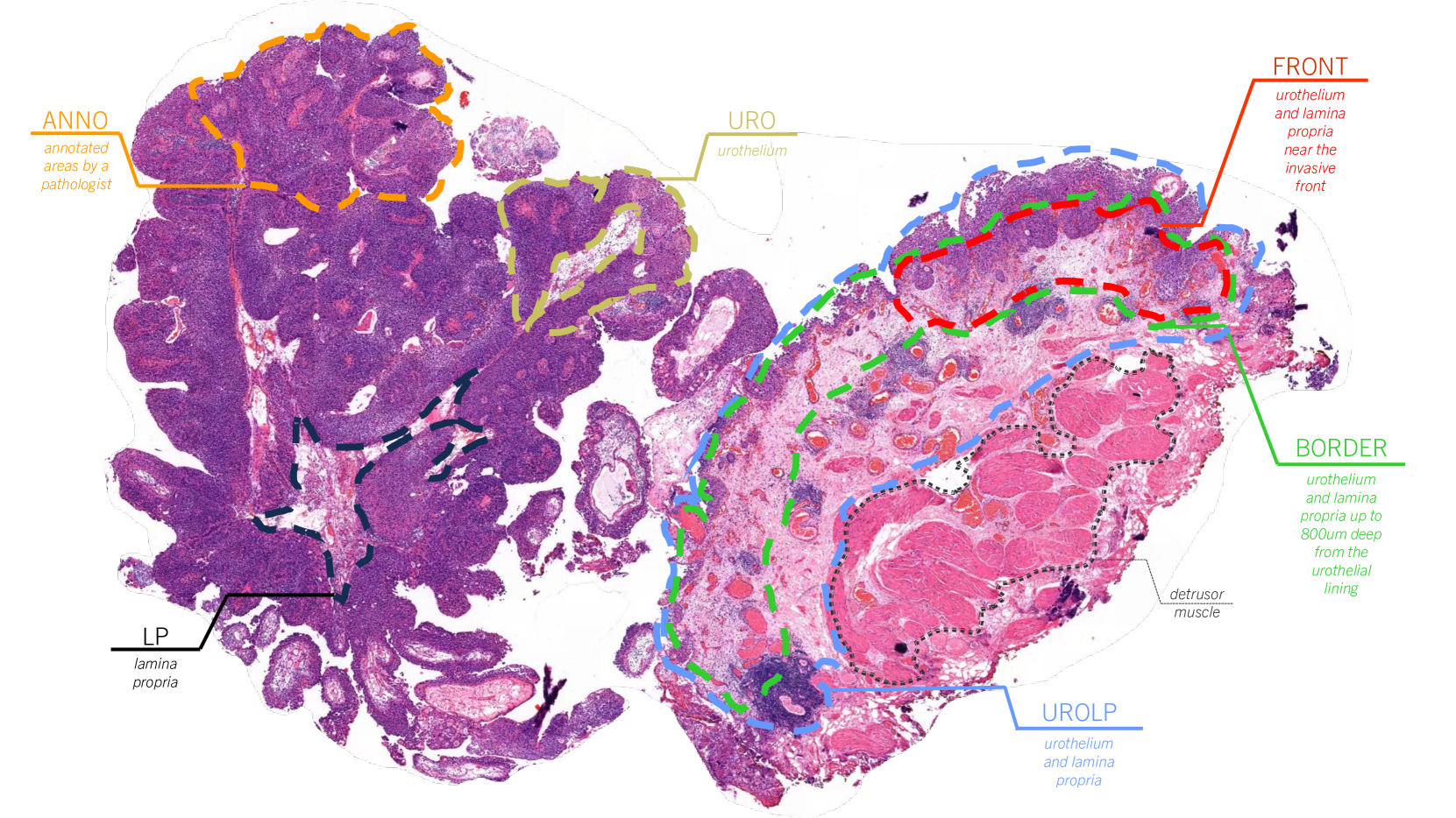
We present a pioneering investigation into the application of deep learning techniques to analyze histopathological images for addressing the substantial challenge of automated prognostic prediction. Prognostic prediction poses a unique challenge as the ground truth labels are inherently weak, and the model must anticipate future events that are not directly observable in the image. To address this challenge, we propose a novel three-part framework comprising of a convolutional network based tissue segmentation algorithm for region of interest delineation, a contrastive learning module for feature extraction, and a nested multiple instance learning classification module. Our study explores the significance of various regions of interest within the histopathological slides and exploits diverse learning scenarios. The pipeline is initially validated on artificially generated data and a simpler diagnostic task. Transitioning to prognostic prediction, tasks become more challenging. Employing bladder cancer as use case, our best models yield an AUC of 0.721 and 0.678 for recurrence and treatment outcome prediction respectively.
Read more5/27/2024
🖼️
0
An interpretable machine learning system for colorectal cancer diagnosis from pathology slides
Pedro C. Neto, Diana Montezuma, Sara P. Oliveira, Domingos Oliveira, Jo~ao Fraga, Ana Monteiro, Jo~ao Monteiro, Liliana Ribeiro, Sofia Gonc{c}alves, Stefan Reinhard, Inti Zlobec, Isabel M. Pinto, Jaime S. Cardoso
Considering the profound transformation affecting pathology practice, we aimed to develop a scalable artificial intelligence (AI) system to diagnose colorectal cancer from whole-slide images (WSI). For this, we propose a deep learning (DL) system that learns from weak labels, a sampling strategy that reduces the number of training samples by a factor of six without compromising performance, an approach to leverage a small subset of fully annotated samples, and a prototype with explainable predictions, active learning features and parallelisation. Noting some problems in the literature, this study is conducted with one of the largest WSI colorectal samples dataset with approximately 10,500 WSIs. Of these samples, 900 are testing samples. Furthermore, the robustness of the proposed method is assessed with two additional external datasets (TCGA and PAIP) and a dataset of samples collected directly from the proposed prototype. Our proposed method predicts, for the patch-based tiles, a class based on the severity of the dysplasia and uses that information to classify the whole slide. It is trained with an interpretable mixed-supervision scheme to leverage the domain knowledge introduced by pathologists through spatial annotations. The mixed-supervision scheme allowed for an intelligent sampling strategy effectively evaluated in several different scenarios without compromising the performance. On the internal dataset, the method shows an accuracy of 93.44% and a sensitivity between positive (low-grade and high-grade dysplasia) and non-neoplastic samples of 0.996. On the external test samples varied with TCGA being the most challenging dataset with an overall accuracy of 84.91% and a sensitivity of 0.996.
Read more5/2/2024
🏷️
0
Classification of Breast Cancer Histopathology Images using a Modified Supervised Contrastive Learning Method
Matina Mahdizadeh Sani, Ali Royat, Mahdieh Soleymani Baghshah
Deep neural networks have reached remarkable achievements in medical image processing tasks, specifically in classifying and detecting various diseases. However, when confronted with limited data, these networks face a critical vulnerability, often succumbing to overfitting by excessively memorizing the limited information available. This work addresses the challenge mentioned above by improving the supervised contrastive learning method leveraging both image-level labels and domain-specific augmentations to enhance model robustness. This approach integrates self-supervised pre-training with a two-stage supervised contrastive learning strategy. In the first stage, we employ a modified supervised contrastive loss that not only focuses on reducing false negatives but also introduces an elimination effect to address false positives. In the second stage, a relaxing mechanism is introduced that refines positive and negative pairs based on similarity, ensuring that only relevant image representations are aligned. We evaluate our method on the BreakHis dataset, which consists of breast cancer histopathology images, and demonstrate an increase in classification accuracy by 1.45% in the image level, compared to the state-of-the-art method. This improvement corresponds to 93.63% absolute accuracy, highlighting the effectiveness of our approach in leveraging properties of data to learn more appropriate representation space.
Read more9/25/2024

0
NMGrad: Advancing Histopathological Bladder Cancer Grading with Weakly Supervised Deep Learning
Saul Fuster, Umay Kiraz, Trygve Eftest{o}l, Emiel A. M. Janssen, Kjersti Engan
The most prevalent form of bladder cancer is urothelial carcinoma, characterized by a high recurrence rate and substantial lifetime treatment costs for patients. Grading is a prime factor for patient risk stratification, although it suffers from inconsistencies and variations among pathologists. Moreover, absence of annotations in medical imaging difficults training deep learning models. To address these challenges, we introduce a pipeline designed for bladder cancer grading using histological slides. First, it extracts urothelium tissue tiles at different magnification levels, employing a convolutional neural network for processing for feature extraction. Then, it engages in the slide-level prediction process. It employs a nested multiple instance learning approach with attention to predict the grade. To distinguish different levels of malignancy within specific regions of the slide, we include the origins of the tiles in our analysis. The attention scores at region level is shown to correlate with verified high-grade regions, giving some explainability to the model. Clinical evaluations demonstrate that our model consistently outperforms previous state-of-the-art methods.
Read more5/27/2024