Comparative Analysis of Image Enhancement Techniques for Brain Tumor Segmentation: Contrast, Histogram, and Hybrid Approaches

0
🖼️
Sign in to get full access
Overview
- This study investigates the impact of image enhancement techniques on Convolutional Neural Network (CNN)-based Brain Tumor Segmentation.
- The researchers focused on Histogram Equalization (HE), Contrast Limited Adaptive Histogram Equalization (CLAHE), and their hybrid variations.
- They used the U-Net architecture on a dataset of 3064 Brain MRI images, exploring preprocessing steps like resizing and enhancement.
- The study provides a detailed analysis of the CNN-based U-Net architecture, training, and validation processes.
- The comparative analysis reveals that the hybrid approach CLAHE-HE consistently outperforms the other techniques.
Plain English Explanation
The researchers in this study wanted to understand how different image enhancement techniques can affect the performance of a type of artificial intelligence (AI) model called a Convolutional Neural Network (CNN) when it comes to identifying and segmenting brain tumors in MRI images. They focused on two specific image enhancement methods: Histogram Equalization (HE) and Contrast Limited Adaptive Histogram Equalization (CLAHE), as well as a hybrid approach that combines the two.
To do this, they used a popular AI architecture called U-Net, which is commonly used for medical image segmentation tasks. They trained and tested the U-Net model on a dataset of 3,064 brain MRI images, experimenting with different preprocessing steps like resizing and applying the image enhancement techniques.
The researchers then compared the performance of the U-Net model across various metrics, such as accuracy, loss, mean squared error (MSE), Intersection over Union (IoU), and Dice Similarity Coefficient (DSC). Their results showed that the hybrid CLAHE-HE approach consistently outperformed the other techniques, achieving very high accuracy and robust segmentation overlap, which is crucial for accurate diagnosis and treatment planning in neuro-oncology.
Technical Explanation
The researchers employed the U-Net architecture, a popular CNN-based segmentation model, on a dataset of 3,064 brain MRI images. They explored the impact of different image enhancement techniques, including Histogram Equalization (HE) and Contrast Limited Adaptive Histogram Equalization (CLAHE), as well as a hybrid approach combining the two.
The study involved a detailed analysis of the U-Net architecture, training, and validation processes. The researchers carefully designed the experiments, including resizing the input images and applying the various enhancement techniques during the preprocessing stage.
The comparative analysis revealed that the hybrid CLAHE-HE approach consistently outperformed the other techniques across several evaluation metrics, such as Accuracy, Loss, MSE, IoU, and DSC. The hybrid method achieved exceptional performance, with training, testing, and validation accuracy of 0.9982, 0.9939, and 0.9936, respectively. The Jaccard (IoU) values were 0.9862, 0.9847, and 0.9864, while the Dice (DSC) values were 0.993, 0.9923, and 0.9932 for the same phases.
These results highlight the potential of the hybrid CLAHE-HE approach in enhancing the accuracy and robustness of brain tumor segmentation, which is crucial for improved diagnosis and treatment planning in neuro-oncology.
Critical Analysis
The study provides a comprehensive investigation into the impact of image enhancement techniques on CNN-based brain tumor segmentation. The researchers thoroughly tested and compared various methods, including the novel hybrid CLAHE-HE approach, which demonstrated impressive performance.
However, the paper does not explicitly address potential limitations or caveats of the study. For instance, the researchers could have explored the generalizability of the findings by testing the models on additional datasets or evaluating their robustness to different tumor types or imaging modalities.
Additionally, the study could have discussed the computational complexity and inference time of the different enhancement techniques, as these factors can be crucial in real-world clinical applications, where efficient and timely processing of medical images is essential.
Further research could also investigate the interpretability and explainability of the CNN-based segmentation models, which could provide valuable insights into the decision-making process and potentially lead to more trustworthy and transparent neuro-oncological applications.
Conclusion
This study systematically explores the impact of image enhancement techniques, particularly Histogram Equalization (HE), Contrast Limited Adaptive Histogram Equalization (CLAHE), and their hybrid variation, on the performance of a CNN-based brain tumor segmentation model. The researchers used the U-Net architecture and a dataset of 3,064 brain MRI images to conduct a detailed analysis.
The results demonstrate that the hybrid CLAHE-HE approach consistently outperforms the other techniques, achieving exceptional accuracy, overlap, and robustness in the segmentation task. These findings highlight the potential of this hybrid method to enhance diagnosis and treatment planning in neuro-oncological applications.
The study's insights contribute to the ongoing efforts to refine segmentation methodologies and improve the precision and reliability of medical image analysis, ultimately benefiting healthcare professionals and patients in the field of neuro-oncology.
This summary was produced with help from an AI and may contain inaccuracies - check out the links to read the original source documents!
Related Papers
🖼️

0
Comparative Analysis of Image Enhancement Techniques for Brain Tumor Segmentation: Contrast, Histogram, and Hybrid Approaches
Shoffan Saifullah, Andri Pranolo, Rafa{l} Dre.zewski
This study systematically investigates the impact of image enhancement techniques on Convolutional Neural Network (CNN)-based Brain Tumor Segmentation, focusing on Histogram Equalization (HE), Contrast Limited Adaptive Histogram Equalization (CLAHE), and their hybrid variations. Employing the U-Net architecture on a dataset of 3064 Brain MRI images, the research delves into preprocessing steps, including resizing and enhancement, to optimize segmentation accuracy. A detailed analysis of the CNN-based U-Net architecture, training, and validation processes is provided. The comparative analysis, utilizing metrics such as Accuracy, Loss, MSE, IoU, and DSC, reveals that the hybrid approach CLAHE-HE consistently outperforms others. Results highlight its superior accuracy (0.9982, 0.9939, 0.9936 for training, testing, and validation, respectively) and robust segmentation overlap, with Jaccard values of 0.9862, 0.9847, and 0.9864, and Dice values of 0.993, 0.9923, and 0.9932 for the same phases, emphasizing its potential in neuro-oncological applications. The study concludes with a call for refinement in segmentation methodologies to further enhance diagnostic precision and treatment planning in neuro-oncology.
Read more4/9/2024
🖼️

0
Exploration of Multi-Scale Image Fusion Systems in Intelligent Medical Image Analysis
Yuxiang Hu, Haowei Yang, Ting Xu, Shuyao He, Jiajie Yuan, Haozhang Deng
The diagnosis of brain cancer relies heavily on medical imaging techniques, with MRI being the most commonly used. It is necessary to perform automatic segmentation of brain tumors on MRI images. This project intends to build an MRI algorithm based on U-Net. The residual network and the module used to enhance the context information are combined, and the void space convolution pooling pyramid is added to the network for processing. The brain glioma MRI image dataset provided by cancer imaging archives was experimentally verified. A multi-scale segmentation method based on a weighted least squares filter was used to complete the 3D reconstruction of brain tumors. Thus, the accuracy of three-dimensional reconstruction is further improved. Experiments show that the local texture features obtained by the proposed algorithm are similar to those obtained by laser scanning. The algorithm is improved by using the U-Net method and an accuracy of 0.9851 is obtained. This approach significantly enhances the precision of image segmentation and boosts the efficiency of image classification.
Read more6/28/2024


0
On Enhancing Brain Tumor Segmentation Across Diverse Populations with Convolutional Neural Networks
Fadillah Maani, Anees Ur Rehman Hashmi, Numan Saeed, Mohammad Yaqub
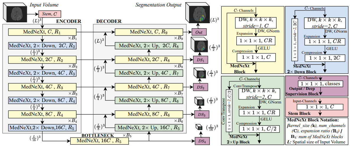
Brain tumor segmentation is a fundamental step in assessing a patient's cancer progression. However, manual segmentation demands significant expert time to identify tumors in 3D multimodal brain MRI scans accurately. This reliance on manual segmentation makes the process prone to intra- and inter-observer variability. This work proposes a brain tumor segmentation method as part of the BraTS-GoAT challenge. The task is to segment tumors in brain MRI scans automatically from various populations, such as adults, pediatrics, and underserved sub-Saharan Africa. We employ a recent CNN architecture for medical image segmentation, namely MedNeXt, as our baseline, and we implement extensive model ensembling and postprocessing for inference. Our experiments show that our method performs well on the unseen validation set with an average DSC of 85.54% and HD95 of 27.88. The code is available on https://github.com/BioMedIA-MBZUAI/BraTS2024_BioMedIAMBZ.
Read more5/7/2024
🤿

0
Deep Learning-Based Brain Image Segmentation for Automated Tumour Detection
Suman Sourabh, Murugappan Valliappan, Narayana Darapaneni, Anwesh R P
Introduction: The present study on the development and evaluation of an automated brain tumor segmentation technique based on deep learning using the 3D U-Net model. Objectives: The objective is to leverage state-of-the-art convolutional neural networks (CNNs) on a large dataset of brain MRI scans for segmentation. Methods: The proposed methodology applies pre-processing techniques for enhanced performance and generalizability. Results: Extensive validation on an independent dataset confirms the model's robustness and potential for integration into clinical workflows. The study emphasizes the importance of data pre-processing and explores various hyperparameters to optimize the model's performance. The 3D U-Net, has given IoUs for training and validation dataset have been 0.8181 and 0.66 respectively. Conclusion: Ultimately, this comprehensive framework showcases the efficacy of deep learning in automating brain tumour detection, offering valuable support in clinical practice.
Read more4/10/2024