CT-based brain ventricle segmentation via diffusion Schrodinger Bridge without target domain ground truths

0

Sign in to get full access
Overview
• This paper presents a novel method for segmenting brain ventricles in computed tomography (CT) images without requiring ground truth segmentation data in the target domain.
• The approach leverages a diffusion-based Schrödinger Bridge model to learn a mapping between source and target domain features, enabling zero-shot segmentation of ventricles in the target CT images.
• The method aims to address the challenge of limited availability of annotated medical imaging data, particularly for rare diseases or new imaging modalities, by enabling knowledge transfer from source to target domains.
Plain English Explanation
In the field of medical image analysis, segmenting brain structures, such as ventricles, is an important task with various clinical applications. However, acquiring the necessary ground truth data (i.e., manually annotated images) for training segmentation models can be a time-consuming and labor-intensive process, especially for rare diseases or new imaging techniques.
To overcome this challenge, the researchers in this paper propose a novel method that can perform brain ventricle segmentation in computed tomography (CT) scans without requiring ground truth segmentation data in the target domain. Instead, their approach relies on a diffusion-based Schrödinger Bridge model to learn a mapping between features in a source domain (e.g., magnetic resonance imaging or MRI) and the target domain (CT).
The key idea is to leverage the availability of annotated data in the source domain (e.g., MRI with ground truth ventricle segmentation) and use the learned feature mapping to perform zero-shot segmentation of ventricles in the target domain (CT). This means that the model can be applied to new CT scans without the need for any manual annotations or segmentation data in the CT domain.
The diffusion-based Schrödinger Bridge model is a powerful technique that can learn the relationship between different data distributions, even in the absence of direct correspondences between them. By using this model, the researchers can effectively transfer knowledge from the source domain to the target domain, enabling accurate segmentation of brain ventricles in CT images without requiring any target-specific ground truth data.
Technical Explanation
The proposed method builds on the diffusion-based Schrödinger Bridge framework, which has been successfully applied to the task of high-quality MRI reconstruction. In this work, the researchers adapt the diffusion Schrödinger Bridge approach to the problem of CT-based brain ventricle segmentation, where the goal is to perform accurate segmentation without any ground truth annotations in the target CT domain.
The key components of the proposed method are:
-
Source Domain Model: The researchers first train a segmentation model on a source domain, such as MRI, where annotated ground truth data is available. This model serves as the initial mapping between the input image and the target ventricle segmentation.
-
Diffusion Schrödinger Bridge: The diffusion Schrödinger Bridge model is then used to learn a transformation between the feature representations of the source (MRI) and target (CT) domains. This allows the source domain segmentation model to be applied to the target CT images, even in the absence of ground truth annotations.
-
Iterative Refinement: The segmentation results on the target CT images are used to iteratively refine the Schrödinger Bridge model, further improving the cross-domain feature alignment and the overall segmentation performance.
The proposed method is evaluated on a dataset of CT brain images, and the results demonstrate that the zero-shot segmentation approach can achieve competitive performance compared to supervised segmentation models trained directly on CT data with ground truth annotations.
Critical Analysis
The authors acknowledge several limitations and areas for further research in their work:
-
Dependence on Source Domain: The performance of the proposed method is inherently dependent on the quality and relevance of the source domain data and model. If the source domain (e.g., MRI) differs significantly from the target domain (CT), the cross-domain feature alignment may be suboptimal, limiting the segmentation accuracy.
-
Scalability to Multiple Domains: The current framework focuses on a single source-target domain pair. Extending the method to handle multiple source domains or more complex domain shifts (e.g., incorporating different imaging modalities or pathologies) may require additional research and model adaptations.
-
Validation on Diverse Datasets: The authors have primarily evaluated their method on a single CT brain image dataset. Further validation on a wider range of CT datasets, including those with different patient populations, scanner types, or imaging protocols, would help demonstrate the broader applicability and robustness of the approach.
-
Interpretability and Explainability: As with many deep learning-based methods, the inner workings of the diffusion Schrödinger Bridge model and its cross-domain feature alignment process may not be fully interpretable. Developing more explainable techniques could enhance the trust and acceptance of such methods in clinical settings.
Despite these limitations, the proposed zero-shot segmentation approach based on the diffusion Schrödinger Bridge model represents an important step towards addressing the challenge of limited annotated data in medical imaging. The ability to leverage knowledge from source domains to perform accurate segmentation in target domains without ground truth annotations has the potential to significantly improve the accessibility and deployability of advanced medical image analysis algorithms in real-world clinical settings.
Conclusion
This paper introduces a novel method for CT-based brain ventricle segmentation that does not require ground truth segmentation data in the target domain. By leveraging a diffusion-based Schrödinger Bridge model, the researchers demonstrate how knowledge can be effectively transferred from a source domain (e.g., MRI) to perform accurate zero-shot segmentation of ventricles in CT images.
The key contribution of this work is the ability to overcome the data annotation challenge that often hinders the development and deployment of medical image analysis algorithms. By enabling segmentation in the absence of ground truth labels, the proposed approach opens up new possibilities for applying advanced deep learning techniques to a wider range of medical imaging applications, particularly in scenarios where annotated data is scarce or difficult to obtain.
The research also highlights the potential of diffusion-based models, such as the Schrödinger Bridge, to facilitate cross-domain knowledge transfer in the medical imaging domain. As the field continues to evolve, further advancements in this area could lead to more robust, scalable, and clinically-deployable medical image analysis tools, ultimately improving patient care and outcomes.
This summary was produced with help from an AI and may contain inaccuracies - check out the links to read the original source documents!
Related Papers
0
CT-based brain ventricle segmentation via diffusion Schrodinger Bridge without target domain ground truths
Reihaneh Teimouri, Marta Kersten-Oertel, Yiming Xiao
Efficient and accurate brain ventricle segmentation from clinical CT scans is critical for emergency surgeries like ventriculostomy. With the challenges in poor soft tissue contrast and a scarcity of well-annotated databases for clinical brain CTs, we introduce a novel uncertainty-aware ventricle segmentation technique without the need of CT segmentation ground truths by leveraging diffusion-model-based domain adaptation. Specifically, our method employs the diffusion Schrodinger Bridge and an attention recurrent residual U-Net to capitalize on unpaired CT and MRI scans to derive automatic CT segmentation from those of the MRIs, which are more accessible. Importantly, we propose an end-to-end, joint training framework of image translation and segmentation tasks, and demonstrate its benefit over training individual tasks separately. By comparing the proposed method against similar setups using two different GAN models for domain adaptation (CycleGAN and CUT), we also reveal the advantage of diffusion models towards improved segmentation and image translation quality. With a Dice score of 0.78$pm$0.27, our proposed method outperformed the compared methods, including SynSeg-Net, while providing intuitive uncertainty measures to further facilitate quality control of the automatic segmentation outcomes. The implementation of our proposed method is available at: https://github.com/HealthX-Lab/DiffusionSynCTSeg.
Read more7/16/2024

0
Label-Efficient 3D Brain Segmentation via Complementary 2D Diffusion Models with Orthogonal Views
Jihoon Cho, Suhyun Ahn, Beomju Kim, Hyungjoon Bae, Xiaofeng Liu, Fangxu Xing, Kyungeun Lee, Georges Elfakhri, Van Wedeen, Jonghye Woo, Jinah Park
Deep learning-based segmentation techniques have shown remarkable performance in brain segmentation, yet their success hinges on the availability of extensive labeled training data. Acquiring such vast datasets, however, poses a significant challenge in many clinical applications. To address this issue, in this work, we propose a novel 3D brain segmentation approach using complementary 2D diffusion models. The core idea behind our approach is to first mine 2D features with semantic information extracted from the 2D diffusion models by taking orthogonal views as input, followed by fusing them into a 3D contextual feature representation. Then, we use these aggregated features to train multi-layer perceptrons to classify the segmentation labels. Our goal is to achieve reliable segmentation quality without requiring complete labels for each individual subject. Our experiments on training in brain subcortical structure segmentation with a dataset from only one subject demonstrate that our approach outperforms state-of-the-art self-supervised learning methods. Further experiments on the minimum requirement of annotation by sparse labeling yield promising results even with only nine slices and a labeled background region.
Read more7/18/2024
0
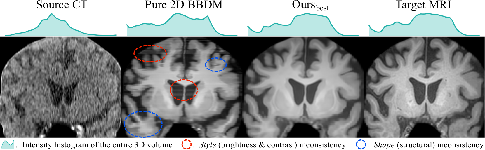
Slice-Consistent 3D Volumetric Brain CT-to-MRI Translation with 2D Brownian Bridge Diffusion Model
Kyobin Choo, Youngjun Jun, Mijin Yun, Seong Jae Hwang
In neuroimaging, generally, brain CT is more cost-effective and accessible imaging option compared to MRI. Nevertheless, CT exhibits inferior soft-tissue contrast and higher noise levels, yielding less precise structural clarity. In response, leveraging more readily available CT to construct its counterpart MRI, namely, medical image-to-image translation (I2I), serves as a promising solution. Particularly, while diffusion models (DMs) have recently risen as a powerhouse, they also come with a few practical caveats for medical I2I. First, DMs' inherent stochasticity from random noise sampling cannot guarantee consistent MRI generation that faithfully reflects its CT. Second, for 3D volumetric images which are prevalent in medical imaging, naively using 2D DMs leads to slice inconsistency, e.g., abnormal structural and brightness changes. While 3D DMs do exist, significant training costs and data dependency bring hesitation. As a solution, we propose novel style key conditioning (SKC) and inter-slice trajectory alignment (ISTA) sampling for the 2D Brownian bridge diffusion model. Specifically, SKC ensures a consistent imaging style (e.g., contrast) across slices, and ISTA interconnects the independent sampling of each slice, deterministically achieving style and shape consistent 3D CT-to-MRI translation. To the best of our knowledge, this study is the first to achieve high-quality 3D medical I2I based only on a 2D DM with no extra architectural models. Our experimental results show superior 3D medical I2I than existing 2D and 3D baselines, using in-house CT-MRI dataset and BraTS2023 FLAIR-T1 MRI dataset.
Read more7/9/2024

0
Integrating Deep Unfolding with Direct Diffusion Bridges for Computed Tomography Reconstruction
Herman Verinaz-Jadan, Su Yan
Computed Tomography (CT) is widely used in healthcare for detailed imaging. However, Low-dose CT, despite reducing radiation exposure, often results in images with compromised quality due to increased noise. Traditional methods, including preprocessing, post-processing, and model-based approaches that leverage physical principles, are employed to improve the quality of image reconstructions from noisy projections or sinograms. Recently, deep learning has significantly advanced the field, with diffusion models outperforming both traditional methods and other deep learning approaches. These models effectively merge deep learning with physics, serving as robust priors for the inverse problem in CT. However, they typically require prolonged computation times during sampling. This paper introduces the first approach to merge deep unfolding with Direct Diffusion Bridges (DDBs) for CT, integrating the physics into the network architecture and facilitating the transition from degraded to clean images by bypassing excessively noisy intermediate stages commonly encountered in diffusion models. Moreover, this approach includes a tailored training procedure that eliminates errors typically accumulated during sampling. The proposed approach requires fewer sampling steps and demonstrates improved fidelity metrics, outperforming many existing state-of-the-art techniques.
Read more9/17/2024