A dataset of primary nasopharyngeal carcinoma MRI with multi-modalities segmentation

0

Sign in to get full access
Overview
- This paper presents a dataset of primary nasopharyngeal carcinoma magnetic resonance imaging (MRI) scans with multi-modal segmentation.
- The dataset includes MRI scans from multiple imaging modalities, along with expert-annotated segmentation of key anatomical regions.
- The dataset aims to facilitate research and development of advanced medical image analysis techniques for the diagnosis and treatment of nasopharyngeal carcinoma.
Plain English Explanation
Nasopharyngeal carcinoma is a type of cancer that develops in the nasopharynx, the upper part of the throat behind the nose. Diagnosing and treating this cancer can be challenging, as it often requires detailed medical imaging and expert analysis.
This research paper introduces a new dataset that can help address this challenge. The dataset contains MRI scans of patients with primary nasopharyngeal carcinoma, along with detailed segmentation (or labeling) of different anatomical structures within the scans. This segmentation was done by medical experts, providing a reliable reference for researchers and clinicians.
By making this dataset publicly available, the authors hope to enable the development of more advanced image analysis techniques that can improve the diagnosis and treatment of nasopharyngeal carcinoma. The dataset can also be used to train deep learning models to automate the segmentation process, potentially saving time and improving consistency.
Overall, this dataset represents an important contribution to the field of medical imaging and cancer diagnosis, as it provides a valuable resource for researchers and clinicians working to better understand and treat nasopharyngeal carcinoma.
Technical Explanation
The paper presents a dataset of 157 primary nasopharyngeal carcinoma patients, each with multi-modal MRI scans (T1-weighted, T2-weighted, and contrast-enhanced T1-weighted) and expert-annotated segmentation of key anatomical structures, including the gross tumor volume, clinical target volume, and organs at risk (such as the brainstem, spinal cord, and parotid glands).
The dataset was collected from a single medical center, with MRI scans acquired using a standardized protocol. The segmentation was performed by experienced radiation oncologists and radiologists, and the accuracy of the annotations was verified through a consensus review process.
The authors provide detailed information on the dataset, including the imaging parameters, segmentation guidelines, and data organization. They also discuss the potential applications of the dataset, such as training deep learning models for automatic segmentation, evaluating the performance of computer-aided diagnosis systems, and developing personalized treatment planning for nasopharyngeal carcinoma patients.
Critical Analysis
The authors acknowledge several limitations of the dataset, including the relatively small sample size, the single-center nature of the data, and the potential for inter-observer variability in the segmentation process. They also note that the dataset only includes primary nasopharyngeal carcinoma cases, and does not cover recurrent or metastatic disease.
Additionally, the dataset does not include clinical outcomes or treatment information, which could limit its utility for certain research applications, such as predicting treatment response or evaluating the impact of image-guided interventions.
Future research could explore ways to expand the dataset, such as by including data from multiple medical centers or incorporating additional clinical and treatment information. Validation of the dataset's utility for various medical imaging tasks would also be valuable.
Conclusion
This dataset of primary nasopharyngeal carcinoma MRI scans with multi-modal segmentation represents an important contribution to the field of medical imaging and cancer research. By making this dataset publicly available, the authors have provided a valuable resource for researchers and clinicians working to develop advanced image analysis techniques and improve the diagnosis and treatment of this challenging disease.
While the dataset has some limitations, it has the potential to significantly advance our understanding of nasopharyngeal carcinoma and support the development of more personalized and effective cancer care.
This summary was produced with help from an AI and may contain inaccuracies - check out the links to read the original source documents!
Related Papers
0
A dataset of primary nasopharyngeal carcinoma MRI with multi-modalities segmentation
Yin Li, Qi Chen, Kai Wang, Meige Li, Liping Si, Yingwei Guo, Yu Xiong, Qixing Wang, Yang Qin, Ling Xu, Patrick van der Smagt, Jun Tang, Nutan Chen
Multi-modality magnetic resonance imaging data with various sequences facilitate the early diagnosis, tumor segmentation, and disease staging in the management of nasopharyngeal carcinoma (NPC). The lack of publicly available, comprehensive datasets limits advancements in diagnosis, treatment planning, and the development of machine learning algorithms for NPC. Addressing this critical need, we introduce the first comprehensive NPC MRI dataset, encompassing MR axial imaging of 277 primary NPC patients. This dataset includes T1-weighted, T2-weighted, and contrast-enhanced T1-weighted sequences, totaling 831 scans. In addition to the corresponding clinical data, manually annotated and labeled segmentations by experienced radiologists offer high-quality data resources from untreated primary NPC.
Read more4/5/2024
🏷️
0
Classification of Nasopharyngeal Cases using DenseNet Deep Learning Architecture
W. S. H. M. W. Ahmad, M. F. A. Fauzi, M. K. Abdullahi, Jenny T. H. Lee, N. S. A. Basry, A Yahaya, A. M. Ismail, A. Adam, Elaine W. L. Chan, F. S. Abas
Nasopharyngeal carcinoma (NPC) is one of the understudied yet deadliest cancers in South East Asia. In Malaysia, the prevalence is identified mainly in Sarawak, among the ethnic of Bidayuh. NPC is often late-diagnosed because it is asymptomatic at the early stage. There are several tissue representations from the nasopharynx biopsy, such as nasopharyngeal inflammation (NPI), lymphoid hyperplasia (LHP), nasopharyngeal carcinoma (NPC) and normal tissue. This paper is our first initiative to identify the difference between NPC, NPI and normal cases. Seven whole slide images (WSIs) with gigapixel resolutions from seven different patients and two hospitals were experimented with using two test setups, consisting of a different set of images. The tissue regions are patched into smaller blocks and classified using DenseNet architecture with 21 dense layers. Two tests are carried out, each for proof of concept (Test 1) and real-test scenario (Test 2). The accuracy achieved for NPC class is 94.8% for Test 1 and 67.0% for Test 2.
Read more4/5/2024
0
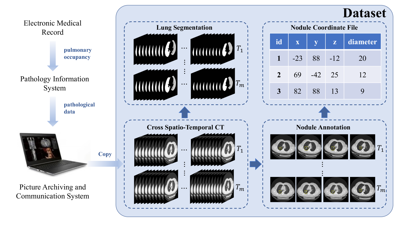
A Cross Spatio-Temporal Pathology-based Lung Nodule Dataset
Muwei Jian, Haoran Zhang, Mingju Shao, Hongyu Chen, Huihui Huang, Yanjie Zhong, Changlei Zhang, Bin Wang, Penghui Gao
Recently, intelligent analysis of lung nodules with the assistant of computer aided detection (CAD) techniques can improve the accuracy rate of lung cancer diagnosis. However, existing CAD systems and pulmonary datasets mainly focus on Computed Tomography (CT) images from one single period, while ignoring the cross spatio-temporal features associated with the progression of nodules contained in imaging data from various captured periods of lung cancer. If the evolution patterns of nodules across various periods in the patients' CT sequences can be explored, it will play a crucial role in guiding the precise screening identification of lung cancer. Therefore, a cross spatio-temporal lung nodule dataset with pathological information for nodule identification and diagnosis is constructed, which contains 328 CT sequences and 362 annotated nodules from 109 patients. This comprehensive database is intended to drive research in the field of CAD towards more practical and robust methods, and also contribute to the further exploration of precision medicine related field. To ensure patient confidentiality, we have removed sensitive information from the dataset.
Read more6/27/2024

0
Exploring Adult Glioma through MRI: A Review of Publicly Available Datasets to Guide Efficient Image Analysis
Meryem Abbad Andaloussi, Raphael Maser, Frank Hertel, Franc{c}ois Lamoline, Andreas Dominik Husch
Publicly available data is essential for the progress of medical image analysis, in particular for crafting machine learning models. Glioma is the most common group of primary brain tumors, and magnetic resonance imaging (MRI) is a widely used modality in their diagnosis and treatment. However, the availability and quality of public datasets for glioma MRI are not well known. In this review, we searched for public datasets for glioma MRI using Google Dataset Search, The Cancer Imaging Archive (TCIA), and Synapse. A total of 28 datasets published between 2005 and May 2024 were found, containing 62019 images from 5515 patients. We analyzed the characteristics of these datasets, such as the origin, size, format, annotation, and accessibility. Additionally, we examined the distribution of tumor types, grades, and stages among the datasets. The implications of the evolution of the WHO classification on tumors of the brain are discussed, in particular the 2021 update that significantly changed the definition of glioblastoma. Additionally, potential research questions that could be explored using these datasets were highlighted, such as tumor evolution through malignant transformation, MRI normalization, and tumor segmentation. Interestingly, only two datasets among the 28 studied reflect the current WHO classification. This review provides a comprehensive overview of the publicly available datasets for glioma MRI currently at our disposal, providing aid to medical image analysis researchers in their decision-making on efficient dataset choice.
Read more9/4/2024