Lensless fiber endomicroscopic phase imaging with speckle-conditioned diffusion model

0

Sign in to get full access
Overview
- Lensless fiber endomicroscopic phase imaging using a speckle-conditioned diffusion model.
- Technique for reconstructing high-quality phase images from speckle patterns captured by a lensless fiber-optic endomicroscope.
- Speckle-conditioned diffusion model enables robust and efficient phase retrieval.
Plain English Explanation
In this research, the scientists developed a new way to take high-quality phase images using a type of microscope called a lensless fiber-optic endomicroscope. This microscope doesn't use any lenses, but instead captures a speckle pattern - a random-looking image made up of bright and dark spots.
The key innovation is a speckle-conditioned diffusion model that can reconstruct a clear phase image from this speckle pattern. Phase images reveal information about the internal structure of the sample being imaged, which is valuable for many biological and medical applications.
The diffusion model essentially "filters out" the random speckle pattern to extract the underlying phase information. This allows the lensless microscope to produce high-quality phase images without needing any bulky or expensive lenses. The technique is efficient and robust, making it well-suited for applications like minimally-invasive medical imaging using small fiber-optic probes.
Technical Explanation
The researchers developed a lensless fiber endomicroscopic imaging system that captures speckle patterns of a sample. These speckle patterns contain information about the phase of the light interacting with the sample, which can be used to reconstruct a phase image.
To enable efficient and robust phase retrieval from the speckle patterns, the researchers introduced a speckle-conditioned diffusion model. This model leverages the statistical properties of speckle patterns to invert the image formation process and recover the underlying phase information.
The diffusion model is trained on simulated speckle patterns, which allows it to learn the relationship between the speckle pattern and the phase of the sample. During inference, the trained model can then rapidly reconstruct the phase image from a captured speckle pattern.
Experiments show that this lensless fiber endomicroscopic phase imaging technique can produce high-quality phase images with good resolution and contrast, without the need for bulky optical components. The simplicity and efficiency of the approach make it well-suited for miniaturized, minimally-invasive imaging applications.
Critical Analysis
The paper provides a compelling demonstration of how advanced computational techniques can enable new imaging modalities with small, simple hardware. The speckle-conditioned diffusion model appears to be a robust and effective way to retrieve phase information from speckle patterns.
However, the authors do acknowledge some limitations of the current system. For example, the field-of-view and resolution are still relatively modest compared to conventional microscopy techniques. There may also be challenges in translating the system to in vivo medical imaging, where factors like tissue scattering and motion could impact performance.
Further research could explore ways to expand the capabilities of the lensless endomicroscope, such as by increasing the field-of-view, improving resolution, or enabling 3D imaging. Investigating the system's sensitivity to practical deployment conditions would also be valuable for assessing its real-world feasibility.
Overall, this work represents an interesting advance in computational imaging that could have important implications for minimally-invasive medical diagnostics and other applications benefiting from compact, lens-free imaging solutions.
Conclusion
This research presents a novel lensless fiber endomicroscopic imaging system that can reconstruct high-quality phase images from speckle patterns using a speckle-conditioned diffusion model. The computational technique enables efficient and robust phase retrieval, overcoming the limitations of traditional lens-based approaches.
The simplicity and compactness of the lensless design make it well-suited for miniaturized, minimally-invasive imaging applications, such as in-vivo medical diagnostics. While the current system has some performance limitations, the underlying computational approach represents an interesting advance in computational imaging that could have broader implications for the field.
This summary was produced with help from an AI and may contain inaccuracies - check out the links to read the original source documents!
Related Papers
0
Lensless fiber endomicroscopic phase imaging with speckle-conditioned diffusion model
Zhaoqing Chen, Jiawei Sun, Xinyi Ye, Bin Zhao, Xuelong Li, Juergen Czarske
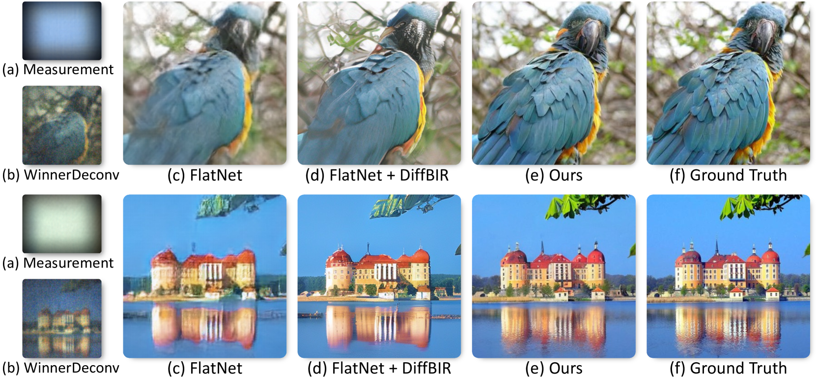
Lensless fiber endomicroscope is an emerging tool for in-vivo microscopic imaging, where quantitative phase imaging (QPI) can be utilized as a label-free method to enhance image contrast. However, existing single-shot phase reconstruction methods through lensless fiber endomicroscope typically perform well on simple images but struggle with complex microscopic structures. Here, we propose a speckle-conditioned diffusion model (SpecDiffusion), which reconstructs phase images directly from speckles captured at the detection side of a multi-core fiber (MCF). Unlike conventional neural networks, SpecDiffusion employs iterative phase denoising steps for speckle-driven phase reconstruction. The iteration scheme allows SpecDiffusion to break down the phase reconstruction process into multiple steps, gradually building up to the final phase image. This attribute alleviates the computation challenge at each step and enables the reconstruction of rich details in complex microscopic images. To validate its efficacy, we build an optical system to capture speckles from MCF and construct a dataset consisting of 100,000 paired images. SpecDiffusion provides high-fidelity phase reconstruction results and shows powerful generalization capacity for unseen objects, such as test charts and biological tissues, reducing the average mean absolute error of the reconstructed tissue images by 7 times. Furthermore, the reconstructed tissue images using SpecDiffusion shows higher accuracy in zero-shot cell segmentation tasks compared to the conventional method, demonstrating the potential for further cell morphology analysis through the learning-based lensless fiber endomicroscope. SpecDiffusion offers a precise and generalized method to phase reconstruction through scattering media, including MCFs, opening new perspective in lensless fiber endomicroscopic imaging.
Read more9/16/2024

0
Single Exposure Quantitative Phase Imaging with a Conventional Microscope using Diffusion Models
Gabriel della Maggiora, Luis Alberto Croquevielle, Harry Horsley, Thomas Heinis, Artur Yakimovich
Phase imaging is gaining importance due to its applications in fields like biomedical imaging and material characterization. In biomedical applications, it can provide quantitative information missing in label-free microscopy modalities. One of the most prominent methods in phase quantification is the Transport-of-Intensity Equation (TIE). TIE often requires multiple acquisitions at different defocus distances, which is not always feasible in a clinical setting. To address this issue, we propose to use chromatic aberrations to induce the required through-focus images with a single exposure, effectively generating a through-focus stack. Since the defocus distance induced by the aberrations is small, conventional TIE solvers are insufficient to address the resulting artifacts. We propose Zero-Mean Diffusion, a modified version of diffusion models designed for quantitative image prediction, and train it with synthetic data to ensure robust phase retrieval. Our contributions offer an alternative TIE approach that leverages chromatic aberrations, achieving accurate single-exposure phase measurement with white light and thus improving the efficiency of phase imaging. Moreover, we present a new class of diffusion models that are well-suited for quantitative data and have a sound theoretical basis. To validate our approach, we employ a widespread brightfield microscope equipped with a commercially available color camera. We apply our model to clinical microscopy of patients' urine, obtaining accurate phase measurements.
Read more6/10/2024
0
PhoCoLens: Photorealistic and Consistent Reconstruction in Lensless Imaging
Xin Cai, Zhiyuan You, Hailong Zhang, Wentao Liu, Jinwei Gu, Tianfan Xue
Lensless cameras offer significant advantages in size, weight, and cost compared to traditional lens-based systems. Without a focusing lens, lensless cameras rely on computational algorithms to recover the scenes from multiplexed measurements. However, current algorithms struggle with inaccurate forward imaging models and insufficient priors to reconstruct high-quality images. To overcome these limitations, we introduce a novel two-stage approach for consistent and photorealistic lensless image reconstruction. The first stage of our approach ensures data consistency by focusing on accurately reconstructing the low-frequency content with a spatially varying deconvolution method that adjusts to changes in the Point Spread Function (PSF) across the camera's field of view. The second stage enhances photorealism by incorporating a generative prior from pre-trained diffusion models. By conditioning on the low-frequency content retrieved in the first stage, the diffusion model effectively reconstructs the high-frequency details that are typically lost in the lensless imaging process, while also maintaining image fidelity. Our method achieves a superior balance between data fidelity and visual quality compared to existing methods, as demonstrated with two popular lensless systems, PhlatCam and DiffuserCam. Project website: https://phocolens.github.io/.
Read more9/27/2024
🧠
0
Pixel super-resolved lensless on-chip sensor with scattering multiplexing
Xuyang Chang, Shaowei Jiang, Yongcun Hu, Liheng Bian
Lensless on-chip microscopy has shown great potential for biomedical imaging due to its large-area and high-throughput imaging capabilities. By combining the pixel super-resolution (PSR) technique, it can improve the resolution beyond the limit of the imaging detector. However, existing PSR techniques are restricted to the feature size and crosstalk of modulation components (such as spatial light modulator), which cannot efficiently encode target information. Besides, the reconstruction algorithms suffer from the trade-off between image quality, reconstruction resolution and computational efficiency. In this work, we constructed a novel integrated lensless on-chip sensor via scattering multiplexing, and reported a robust PSR algorithm for sample reconstruction. The sensor employed a scattering layer as a modulator, which was permanently integrated with the detector. Benefiting from the high-degree-of-freedom reconstruction of the scattering layer, we realized fine wavefront modulation with a small feature size. The integration engineering avoided repetitious calibration and reduce the measurement complexity. The reported PSR algorithm combines both model-driven and data-driven strategies to efficiently exploit the high-frequency information from the fine modulation. A series of experiments validated that the reported sensor provides a low-cost solution for large-scale microscopic imaging, with significant advantages in resolution, image contrast and noise robustness.
Read more9/6/2024