EFCNet: Every Feature Counts for Small Medical Object Segmentation

0

Sign in to get full access
Overview
- This paper introduces EFCNet, a new deep learning model for small medical object segmentation.
- EFCNet aims to improve the performance of medical image segmentation, particularly for small objects that can be challenging to detect.
- The key idea is to leverage every available feature in the network to enhance the segmentation of small objects.
Plain English Explanation
Medical image analysis is an important task in healthcare, as it allows doctors to accurately identify and analyze various structures in the body. One particularly challenging aspect of this is segmenting small objects, such as tiny tumors or lesions, which can be difficult for AI models to detect accurately.
The EFCNet model proposed in this paper tackles this problem by making sure that every feature learned by the network is used to help with the segmentation task, especially for those small, hard-to-find objects. The researchers designed the network architecture and training process to ensure that all available information is leveraged to segment these tiny medical structures as precisely as possible.
By focusing on extracting and using all relevant features, rather than just the most obvious ones, the EFCNet model is able to achieve stronger performance on small medical object segmentation compared to other approaches. This could lead to more accurate diagnosis and treatment planning for patients, as doctors would be able to identify even the smallest abnormalities in medical scans.
Technical Explanation
The key innovation in the EFCNet model is its architecture, which is designed to make the most of all the visual features learned by the network. Rather than just relying on the high-level features extracted by the later layers, EFCNet also incorporates lower-level features from earlier layers to better capture small details.
This is achieved through a series of feature fusion modules that combine information from multiple levels of the network. By aggregating features across scales, EFCNet can assemble a more comprehensive representation that is well-suited for segmenting small objects.
The researchers also developed a novel training strategy that encourages the model to pay attention to small regions during the learning process. This helps EFCNet develop a stronger sensitivity to subtle patterns and boundaries, which is crucial for segmenting tiny medical structures accurately.
Critical Analysis
The EFCNet paper makes a compelling case for the importance of leveraging all available features when tackling small object segmentation in medical imaging. The proposed architecture and training approach show promising results, outperforming other state-of-the-art models on relevant benchmark tasks.
That said, the paper does not extensively explore the limitations of the EFCNet approach. For instance, it would be helpful to understand how the model's performance scales as the size of the target objects decreases further. Additionally, the generalization of EFCNet to a wider range of medical imaging modalities and clinical applications could be examined in future work.
Overall, the EFCNet research represents a valuable contribution to the field of medical image analysis. By emphasizing the importance of comprehensive feature learning, this work opens up new directions for developing even more accurate and reliable AI-powered tools for medical diagnosis and treatment.
Conclusion
The EFCNet model introduced in this paper addresses the challenge of small medical object segmentation by ensuring that every available feature in the network is leveraged for the task. Through its innovative architecture and training strategy, EFCNet is able to outperform other state-of-the-art approaches on relevant benchmarks.
This research highlights the significance of comprehensive feature learning for tackling difficult computer vision problems, particularly in the medical domain where the accurate identification of small structures can be crucial for patient care. While the paper does not fully explore the limitations of the EFCNet approach, it represents an important step forward in developing more robust and reliable AI-based medical image analysis tools.
This summary was produced with help from an AI and may contain inaccuracies - check out the links to read the original source documents!
Related Papers
0
EFCNet: Every Feature Counts for Small Medical Object Segmentation
Lingjie Kong, Qiaoling Wei, Chengming Xu, Han Chen, Yanwei Fu
This paper explores the segmentation of very small medical objects with significant clinical value. While Convolutional Neural Networks (CNNs), particularly UNet-like models, and recent Transformers have shown substantial progress in image segmentation, our empirical findings reveal their poor performance in segmenting the small medical objects and lesions concerned in this paper. This limitation may be attributed to information loss during their encoding and decoding process. In response to this challenge, we propose a novel model named EFCNet for small object segmentation in medical images. Our model incorporates two modules: the Cross-Stage Axial Attention Module (CSAA) and the Multi-Precision Supervision Module (MPS). These modules address information loss during encoding and decoding procedures, respectively. Specifically, CSAA integrates features from all stages of the encoder to adaptively learn suitable information needed in different decoding stages, thereby reducing information loss in the encoder. On the other hand, MPS introduces a novel multi-precision supervision mechanism to the decoder. This mechanism prioritizes attention to low-resolution features in the initial stages of the decoder, mitigating information loss caused by subsequent convolution and sampling processes and enhancing the model's global perception. We evaluate our model on two benchmark medical image datasets. The results demonstrate that EFCNet significantly outperforms previous segmentation methods designed for both medical and normal images.
Read more6/27/2024

0
Spatial-Frequency Dual Progressive Attention Network For Medical Image Segmentation
Zhenhuan Zhou, Along He, Yanlin Wu, Rui Yao, Xueshuo Xie, Tao Li
In medical images, various types of lesions often manifest significant differences in their shape and texture. Accurate medical image segmentation demands deep learning models with robust capabilities in multi-scale and boundary feature learning. However, previous networks still have limitations in addressing the above issues. Firstly, previous networks simultaneously fuse multi-level features or employ deep supervision to enhance multi-scale learning. However, this may lead to feature redundancy and excessive computational overhead, which is not conducive to network training and clinical deployment. Secondly, the majority of medical image segmentation networks exclusively learn features in the spatial domain, disregarding the abundant global information in the frequency domain. This results in a bias towards low-frequency components, neglecting crucial high-frequency information. To address these problems, we introduce SF-UNet, a spatial-frequency dual-domain attention network. It comprises two main components: the Multi-scale Progressive Channel Attention (MPCA) block, which progressively extract multi-scale features across adjacent encoder layers, and the lightweight Frequency-Spatial Attention (FSA) block, with only 0.05M parameters, enabling concurrent learning of texture and boundary features from both spatial and frequency domains. We validate the effectiveness of the proposed SF-UNet on three public datasets. Experimental results show that compared to previous state-of-the-art (SOTA) medical image segmentation networks, SF-UNet achieves the best performance, and achieves up to 9.4% and 10.78% improvement in DSC and IOU. Codes will be released at https://github.com/nkicsl/SF-UNet.
Read more8/20/2024

0
SvANet: A Scale-variant Attention-based Network for Small Medical Object Segmentation
Wei Dai, Rui Liu, Zixuan Wu, Tianyi Wu, Min Wang, Junxian Zhou, Yixuan Yuan, Jun Liu
Early detection and accurate diagnosis can predict the risk of malignant disease transformation, thereby increasing the probability of effective treatment. Identifying mild syndrome with small pathological regions serves as an ominous warning and is fundamental in the early diagnosis of diseases. While deep learning algorithms, particularly convolutional neural networks (CNNs), have shown promise in segmenting medical objects, analyzing small areas in medical images remains challenging. This difficulty arises due to information losses and compression defects from convolution and pooling operations in CNNs, which become more pronounced as the network deepens, especially for small medical objects. To address these challenges, we propose a novel scale-variant attention-based network (SvANet) for accurately segmenting small-scale objects in medical images. The SvANet consists of scale-variant attention, cross-scale guidance, Monte Carlo attention, and vision transformer, which incorporates cross-scale features and alleviates compression artifacts for enhancing the discrimination of small medical objects. Quantitative experimental results demonstrate the superior performance of SvANet, achieving 96.12%, 96.11%, 89.79%, 84.15%, 80.25%, 73.05%, and 72.58% in mean Dice coefficient for segmenting kidney tumors, skin lesions, hepatic tumors, polyps, surgical excision cells, retinal vasculatures, and sperms, which occupy less than 1% of the image areas in KiTS23, ISIC 2018, ATLAS, PolypGen, TissueNet, FIVES, and SpermHealth datasets, respectively.
Read more8/6/2024
0
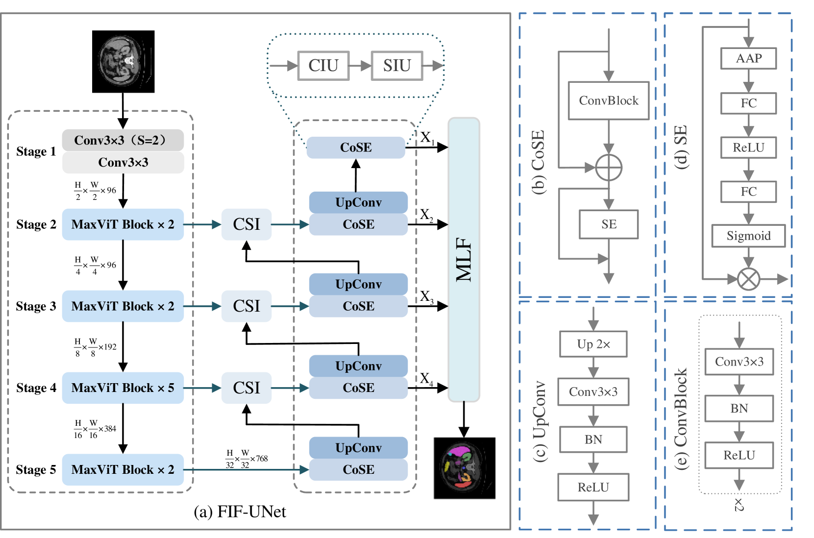
FIF-UNet: An Efficient UNet Using Feature Interaction and Fusion for Medical Image Segmentation
Xiaolin Gou, Chuanlin Liao, Jizhe Zhou, Fengshuo Ye, Yi Lin
Nowadays, pre-trained encoders are widely used in medical image segmentation because of their ability to capture complex feature representations. However, the existing models fail to effectively utilize the rich features obtained by the pre-trained encoder, resulting in suboptimal segmentation results. In this work, a novel U-shaped model, called FIF-UNet, is proposed to address the above issue, including three plug-and-play modules. A channel spatial interaction module (CSI) is proposed to obtain informative features by establishing the interaction between encoder stages and corresponding decoder stages. A cascaded conv-SE module (CoSE) is designed to enhance the representation of critical features by adaptively assigning importance weights on different feature channels. A multi-level fusion module (MLF) is proposed to fuse the multi-scale features from the decoder stages, ensuring accurate and robust final segmentation. Comprehensive experiments on the Synapse and ACDC datasets demonstrate that the proposed FIF-UNet outperforms existing state-of-the-art methods, which achieves the highest average DICE of 86.05% and 92.58%, respectively.
Read more9/10/2024