EXACT-Net:EHR-guided lung tumor auto-segmentation for non-small cell lung cancer radiotherapy

0

Sign in to get full access
Overview
- EXACT-Net is a deep learning model for automatically segmenting lung tumors in non-small cell lung cancer patients.
- The model uses information from electronic health records (EHRs) to guide the segmentation process.
- Accurate tumor segmentation is crucial for delivering precise radiotherapy treatments.
Plain English Explanation
The paper describes a new deep learning model called EXACT-Net that can automatically segment, or outline, lung tumors in non-small cell lung cancer patients. This is an important step for delivering effective radiation therapy, as it allows doctors to target the tumor precisely while avoiding healthy surrounding tissue.
What makes EXACT-Net unique is that it incorporates information from the patient's electronic health records (EHRs) to guide the segmentation process. EHRs contain detailed medical data that can provide valuable context to help the model better identify the tumor boundaries.
By using this EHR data, EXACT-Net aims to improve upon previous lung tumor segmentation models that relied solely on medical images. The researchers tested the model on a large dataset of lung cancer patients and found that it was able to segment the tumors more accurately compared to other state-of-the-art methods.
Improving the accuracy of lung tumor segmentation is critical for ensuring radiation treatments are as effective and targeted as possible. EXACT-Net represents an important step forward in applying advanced AI techniques to this clinical challenge.
Technical Explanation
The researchers developed EXACT-Net, a deep learning model for lung tumor auto-segmentation in non-small cell lung cancer patients. EXACT-Net integrates information from a patient's EHR data, such as clinical notes and laboratory results, to guide the segmentation of tumors in CT scans.
The model architecture consists of a multi-input encoder-decoder network. One input branch takes in the CT images, while the other ingests the structured EHR data. These inputs are processed through separate encoding pathways before being fused and passed through a decoding network to produce the final segmentation.
The researchers evaluated EXACT-Net on a large dataset of lung cancer patients, comparing its performance to other state-of-the-art segmentation models that use only image data. They found that incorporating the EHR information allowed EXACT-Net to achieve significantly higher segmentation accuracy, as measured by common evaluation metrics.
Critical Analysis
The paper provides a compelling demonstration of how leveraging EHR data can enhance the performance of medical image analysis models like EXACT-Net. Integrating diverse data sources is an important frontier in developing robust and clinically-relevant AI systems for healthcare.
However, the authors acknowledge several limitations of their work. The model was trained and evaluated on a single institutional dataset, so further validation on external patient cohorts would be necessary to assess its generalizability. Additionally, the interpretability of the EHR-based features used by the model is not deeply explored, which could limit clinician trust and adoption.
Future research could investigate techniques to better explain the contributions of the EHR data, as well as explore ways to make the model more robust to variations in data availability and quality across healthcare providers. Addressing these challenges could help drive wider adoption of EHR-guided AI systems like EXACT-Net in clinical practice.
Conclusion
The EXACT-Net model demonstrates how incorporating electronic health record data can enhance the performance of deep learning-based lung tumor segmentation. By leveraging this rich source of patient information, the model was able to achieve superior accuracy compared to image-only approaches.
This work highlights the potential of multi-modal AI systems that fuse diverse data sources to tackle complex medical imaging and diagnosis tasks. As hospitals and health systems continue to digitize patient records, techniques like those used in EXACT-Net could become increasingly valuable for delivering precise, personalized cancer care.
While further research is needed to address the model's limitations, this paper represents an important step forward in applying advanced AI to the challenge of automating and improving radiotherapy planning for non-small cell lung cancer.
This summary was produced with help from an AI and may contain inaccuracies - check out the links to read the original source documents!
Related Papers
0
EXACT-Net:EHR-guided lung tumor auto-segmentation for non-small cell lung cancer radiotherapy
Hamed Hooshangnejad, Xue Feng, Gaofeng Huang, Rui Zhang, Katelyn Kelly, Quan Chen, Kai Ding
Lung cancer is a devastating disease with the highest mortality rate among cancer types. Over 60% of non-small cell lung cancer (NSCLC) patients, which accounts for 87% of diagnoses, require radiation therapy. Rapid treatment initiation significantly increases the patient's survival rate and reduces the mortality rate. Accurate tumor segmentation is a critical step in the diagnosis and treatment of NSCLC. Manual segmentation is time and labor-consuming and causes delays in treatment initiation. Although many lung nodule detection methods, including deep learning-based models, have been proposed, there is still a long-standing problem of high false positives (FPs) with most of these methods. Here, we developed an electronic health record (EHR) guided lung tumor auto-segmentation called EXACT-Net (EHR-enhanced eXACtitude in Tumor segmentation), where the extracted information from EHRs using a pre-trained large language model (LLM), was used to remove the FPs and keep the TP nodules only. The auto-segmentation model was trained on NSCLC patients' computed tomography (CT), and the pre-trained LLM was used with the zero-shot learning approach. Our approach resulted in a 250% boost in successful nodule detection using the data from ten NSCLC patients treated in our institution.
Read more8/2/2024

0
Segmentation of Non-Small Cell Lung Carcinomas: Introducing DRU-Net and Multi-Lens Distortion
Soroush Oskouei, Marit Valla, Andr'e Pedersen, Erik Smistad, Vibeke Grotnes Dale, Maren H{o}ib{o}, Sissel Gyrid Freim Wahl, Mats Dehli Haugum, Thomas Lang{o}, Maria Paula Ramnefjell, Lars Andreas Akslen, Gabriel Kiss, Hanne Sorger
Considering the increased workload in pathology laboratories today, automated tools such as artificial intelligence models can help pathologists with their tasks and ease the workload. In this paper, we are proposing a segmentation model (DRU-Net) that can provide a delineation of human non-small cell lung carcinomas and an augmentation method that can improve classification results. The proposed model is a fused combination of truncated pre-trained DenseNet201 and ResNet101V2 as a patch-wise classifier followed by a lightweight U-Net as a refinement model. We have used two datasets (Norwegian Lung Cancer Biobank and Haukeland University Hospital lung cancer cohort) to create our proposed model. The DRU-Net model achieves an average of 0.91 Dice similarity coefficient. The proposed spatial augmentation method (multi-lens distortion) improved the network performance by 3%. Our findings show that choosing image patches that specifically include regions of interest leads to better results for the patch-wise classifier compared to other sampling methods. The qualitative analysis showed that the DRU-Net model is generally successful in detecting the tumor. On the test set, some of the cases showed areas of false positive and false negative segmentation in the periphery, particularly in tumors with inflammatory and reactive changes.
Read more6/21/2024
0
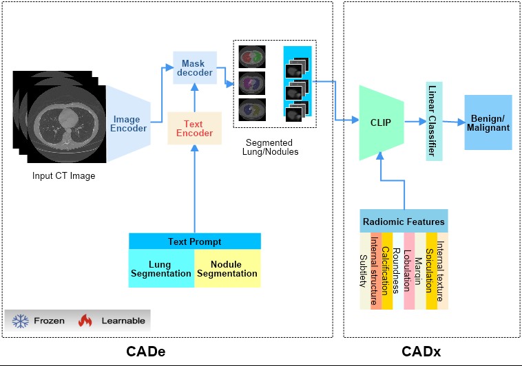
Lung-CADex: Fully automatic Zero-Shot Detection and Classification of Lung Nodules in Thoracic CT Images
Furqan Shaukat, Syed Muhammad Anwar, Abhijeet Parida, Van Khanh Lam, Marius George Linguraru, Mubarak Shah
Lung cancer has been one of the major threats to human life for decades. Computer-aided diagnosis can help with early lung nodul detection and facilitate subsequent nodule characterization. Large Visual Language models (VLMs) have been found effective for multiple downstream medical tasks that rely on both imaging and text data. However, lesion level detection and subsequent diagnosis using VLMs have not been explored yet. We propose CADe, for segmenting lung nodules in a zero-shot manner using a variant of the Segment Anything Model called MedSAM. CADe trains on a prompt suite on input computed tomography (CT) scans by using the CLIP text encoder through prefix tuning. We also propose, CADx, a method for the nodule characterization as benign/malignant by making a gallery of radiomic features and aligning image-feature pairs through contrastive learning. Training and validation of CADe and CADx have been done using one of the largest publicly available datasets, called LIDC. To check the generalization ability of the model, it is also evaluated on a challenging dataset, LUNGx. Our experimental results show that the proposed methods achieve a sensitivity of 0.86 compared to 0.76 that of other fully supervised methods.The source code, datasets and pre-processed data can be accessed using the link:
Read more7/4/2024

0
Deep Learning-Based Segmentation of Tumors in PET/CT Volumes: Benchmark of Different Architectures and Training Strategies
Monika G'orka, Daniel Jaworek, Marek Wodzinski
Cancer is one of the leading causes of death globally, and early diagnosis is crucial for patient survival. Deep learning algorithms have great potential for automatic cancer analysis. Artificial intelligence has achieved high performance in recognizing and segmenting single lesions. However, diagnosing multiple lesions remains a challenge. This study examines and compares various neural network architectures and training strategies for automatically segmentation of cancer lesions using PET/CT images from the head, neck, and whole body. The authors analyzed datasets from the AutoPET and HECKTOR challenges, exploring popular single-step segmentation architectures and presenting a two-step approach. The results indicate that the V-Net and nnU-Net models were the most effective for their respective datasets. The results for the HECKTOR dataset ranged from 0.75 to 0.76 for the aggregated Dice coefficient. Eliminating cancer-free cases from the AutoPET dataset was found to improve the performance of most models. In the case of AutoPET data, the average segmentation efficiency after training only on images containing cancer lesions increased from 0.55 to 0.66 for the classic Dice coefficient and from 0.65 to 0.73 for the aggregated Dice coefficient. The research demonstrates the potential of artificial intelligence in precise oncological diagnostics and may contribute to the development of more targeted and effective cancer assessment techniques.
Read more4/16/2024