A Flexible 2.5D Medical Image Segmentation Approach with In-Slice and Cross-Slice Attention

0
Sign in to get full access
Overview
• This paper introduces a flexible 2.5D medical image segmentation approach that combines in-slice and cross-slice attention mechanisms to enhance the model's ability to capture both local and global spatial features.
• The proposed method, called 2.5D-Attention, aims to address the limitations of existing 2D and 3D segmentation models by leveraging the advantages of both approaches.
• The authors demonstrate the effectiveness of their method on several medical image segmentation tasks, including brain tumor, heart, and liver segmentation, and show that it outperforms state-of-the-art 2D and 3.5D (hybrid 2D-3D) models.
Plain English Explanation
The paper presents a new way to segment, or separate, different parts of medical images, such as MRI scans of the brain, heart, or liver. Existing methods either look at each 2D slice of the image independently or try to incorporate 3D information, but the authors argue that their approach, called 2.5D-Attention, is more flexible and effective.
The key idea is to use attention mechanisms that can focus on both the details within each 2D slice of the image and the relationships between adjacent slices. This allows the model to capture both local and global spatial features, which is important for accurately segmenting complex medical structures.
The authors test their method on several medical imaging tasks and show that it outperforms other state-of-the-art approaches. This suggests that the 2.5D-Attention method could be a valuable tool for medical image analysis and diagnosis.
Technical Explanation
The authors propose a 2.5D medical image segmentation approach that combines in-slice and cross-slice attention mechanisms to enhance the model's ability to capture both local and global spatial features. The in-slice attention module focuses on learning the spatial relationships within each 2D slice, while the cross-slice attention module learns the dependencies between adjacent slices.
The overall architecture of the 2.5D-Attention model consists of an encoder-decoder structure, where the encoder extracts multi-scale features from the input image and the decoder progressively refines the segmentation output. The in-slice and cross-slice attention modules are integrated into the encoder to selectively attend to relevant spatial and channel-wise features.
The authors evaluate their method on several medical image segmentation tasks, including brain tumor, heart, and liver segmentation, using publicly available datasets. They compare the performance of 2.5D-Attention to state-of-the-art 2D and 3.5D (hybrid 2D-3D) models and demonstrate that their approach outperforms these methods in terms of segmentation accuracy and other key metrics.
Critical Analysis
The authors acknowledge that their method still has some limitations, such as the potential for increased computational complexity due to the additional attention modules. They also suggest that further research is needed to explore the application of their approach to other medical imaging modalities and tasks.
Additionally, while the 2.5D-Attention model demonstrates strong performance on the tested medical image segmentation tasks, it would be valuable to see how it compares to more recent 3D-focused methods, such as SegFormer3D, Closer Look at Spatial-Slice Features Learning, and Agile3D, which may offer additional performance improvements or computational efficiency.
Conclusion
The proposed 2.5D-Attention method represents a flexible and effective approach to medical image segmentation that leverages both local and global spatial features. By integrating in-slice and cross-slice attention mechanisms, the model is able to outperform state-of-the-art 2D and 3.5D segmentation techniques on several challenging medical imaging tasks.
While the method has some limitations, it offers a promising direction for further research and development in the field of medical image analysis. The insights and techniques presented in this paper could inspire the design of even more advanced segmentation models that can support improved disease diagnosis, treatment planning, and patient monitoring.
This summary was produced with help from an AI and may contain inaccuracies - check out the links to read the original source documents!
Related Papers
0
A Flexible 2.5D Medical Image Segmentation Approach with In-Slice and Cross-Slice Attention
Amarjeet Kumar, Hongxu Jiang, Muhammad Imran, Cyndi Valdes, Gabriela Leon, Dahyun Kang, Parvathi Nataraj, Yuyin Zhou, Michael D. Weiss, Wei Shao
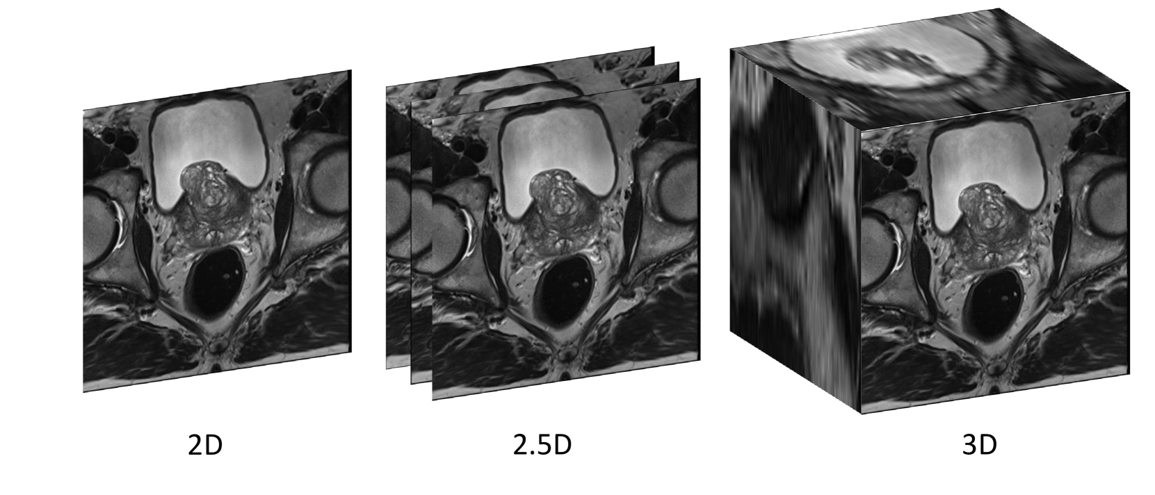
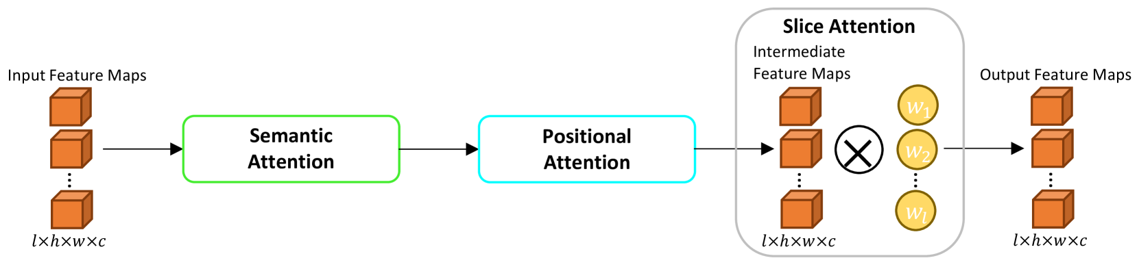
Deep learning has become the de facto method for medical image segmentation, with 3D segmentation models excelling in capturing complex 3D structures and 2D models offering high computational efficiency. However, segmenting 2.5D images, which have high in-plane but low through-plane resolution, is a relatively unexplored challenge. While applying 2D models to individual slices of a 2.5D image is feasible, it fails to capture the spatial relationships between slices. On the other hand, 3D models face challenges such as resolution inconsistencies in 2.5D images, along with computational complexity and susceptibility to overfitting when trained with limited data. In this context, 2.5D models, which capture inter-slice correlations using only 2D neural networks, emerge as a promising solution due to their reduced computational demand and simplicity in implementation. In this paper, we introduce CSA-Net, a flexible 2.5D segmentation model capable of processing 2.5D images with an arbitrary number of slices through an innovative Cross-Slice Attention (CSA) module. This module uses the cross-slice attention mechanism to effectively capture 3D spatial information by learning long-range dependencies between the center slice (for segmentation) and its neighboring slices. Moreover, CSA-Net utilizes the self-attention mechanism to understand correlations among pixels within the center slice. We evaluated CSA-Net on three 2.5D segmentation tasks: (1) multi-class brain MRI segmentation, (2) binary prostate MRI segmentation, and (3) multi-class prostate MRI segmentation. CSA-Net outperformed leading 2D and 2.5D segmentation methods across all three tasks, demonstrating its efficacy and superiority. Our code is publicly available at https://github.com/mirthAI/CSA-Net.
Read more5/2/2024
0
Cross-Slice Attention and Evidential Critical Loss for Uncertainty-Aware Prostate Cancer Detection
Alex Ling Yu Hung, Haoxin Zheng, Kai Zhao, Kaifeng Pang, Demetri Terzopoulos, Kyunghyun Sung
Current deep learning-based models typically analyze medical images in either 2D or 3D albeit disregarding volumetric information or suffering sub-optimal performance due to the anisotropic resolution of MR data. Furthermore, providing an accurate uncertainty estimation is beneficial to clinicians, as it indicates how confident a model is about its prediction. We propose a novel 2.5D cross-slice attention model that utilizes both global and local information, along with an evidential critical loss, to perform evidential deep learning for the detection in MR images of prostate cancer, one of the most common cancers and a leading cause of cancer-related death in men. We perform extensive experiments with our model on two different datasets and achieve state-of-the-art performance in prostate cancer detection along with improved epistemic uncertainty estimation. The implementation of the model is available at https://github.com/aL3x-O-o-Hung/GLCSA_ECLoss.
Read more7/2/2024

0
MSA2Net: Multi-scale Adaptive Attention-guided Network for Medical Image Segmentation
Sina Ghorbani Kolahi, Seyed Kamal Chaharsooghi, Toktam Khatibi, Afshin Bozorgpour, Reza Azad, Moein Heidari, Ilker Hacihaliloglu, Dorit Merhof
Medical image segmentation involves identifying and separating object instances in a medical image to delineate various tissues and structures, a task complicated by the significant variations in size, shape, and density of these features. Convolutional neural networks (CNNs) have traditionally been used for this task but have limitations in capturing long-range dependencies. Transformers, equipped with self-attention mechanisms, aim to address this problem. However, in medical image segmentation it is beneficial to merge both local and global features to effectively integrate feature maps across various scales, capturing both detailed features and broader semantic elements for dealing with variations in structures. In this paper, we introduce MSA$^2$Net, a new deep segmentation framework featuring an expedient design of skip-connections. These connections facilitate feature fusion by dynamically weighting and combining coarse-grained encoder features with fine-grained decoder feature maps. Specifically, we propose a Multi-Scale Adaptive Spatial Attention Gate (MASAG), which dynamically adjusts the receptive field (Local and Global contextual information) to ensure that spatially relevant features are selectively highlighted while minimizing background distractions. Extensive evaluations involving dermatology, and radiological datasets demonstrate that our MSA$^2$Net outperforms state-of-the-art (SOTA) works or matches their performance. The source code is publicly available at https://github.com/xmindflow/MSA-2Net.
Read more8/6/2024

0
Enhancing Single-Slice Segmentation with 3D-to-2D Unpaired Scan Distillation
Xin Yu, Qi Yang, Han Liu, Ho Hin Lee, Yucheng Tang, Lucas W. Remedios, Michael E. Kim, Rendong Zhang, Shunxing Bao, Yuankai Huo, Ann Zenobia Moore, Luigi Ferrucci, Bennett A. Landman
2D single-slice abdominal computed tomography (CT) enables the assessment of body habitus and organ health with low radiation exposure. However, single-slice data necessitates the use of 2D networks for segmentation, but these networks often struggle to capture contextual information effectively. Consequently, even when trained on identical datasets, 3D networks typically achieve superior segmentation results. In this work, we propose a novel 3D-to-2D distillation framework, leveraging pre-trained 3D models to enhance 2D single-slice segmentation. Specifically, we extract the prediction distribution centroid from the 3D representations, to guide the 2D student by learning intra- and inter-class correlation. Unlike traditional knowledge distillation methods that require the same data input, our approach employs unpaired 3D CT scans with any contrast to guide the 2D student model. Experiments conducted on 707 subjects from the single-slice Baltimore Longitudinal Study of Aging (BLSA) dataset demonstrate that state-of-the-art 2D multi-organ segmentation methods can benefit from the 3D teacher model, achieving enhanced performance in single-slice multi-organ segmentation. Notably, our approach demonstrates considerable efficacy in low-data regimes, outperforming the model trained with all available training subjects even when utilizing only 200 training subjects. Thus, this work underscores the potential to alleviate manual annotation burdens.
Read more7/15/2024