Full-Scale Indexing and Semantic Annotation of CT Imaging: Boosting FAIRness

0
🚀
Sign in to get full access
Overview
- The paper explores integrating artificial intelligence (AI) into medical imaging, specifically computed tomography (CT) scans, to improve data management and enable more advanced healthcare applications.
- The proposed approach focuses on semantically enhancing CT image series using automated indexing and segmentation techniques to improve findability, accessibility, interoperability, and reusability of the data.
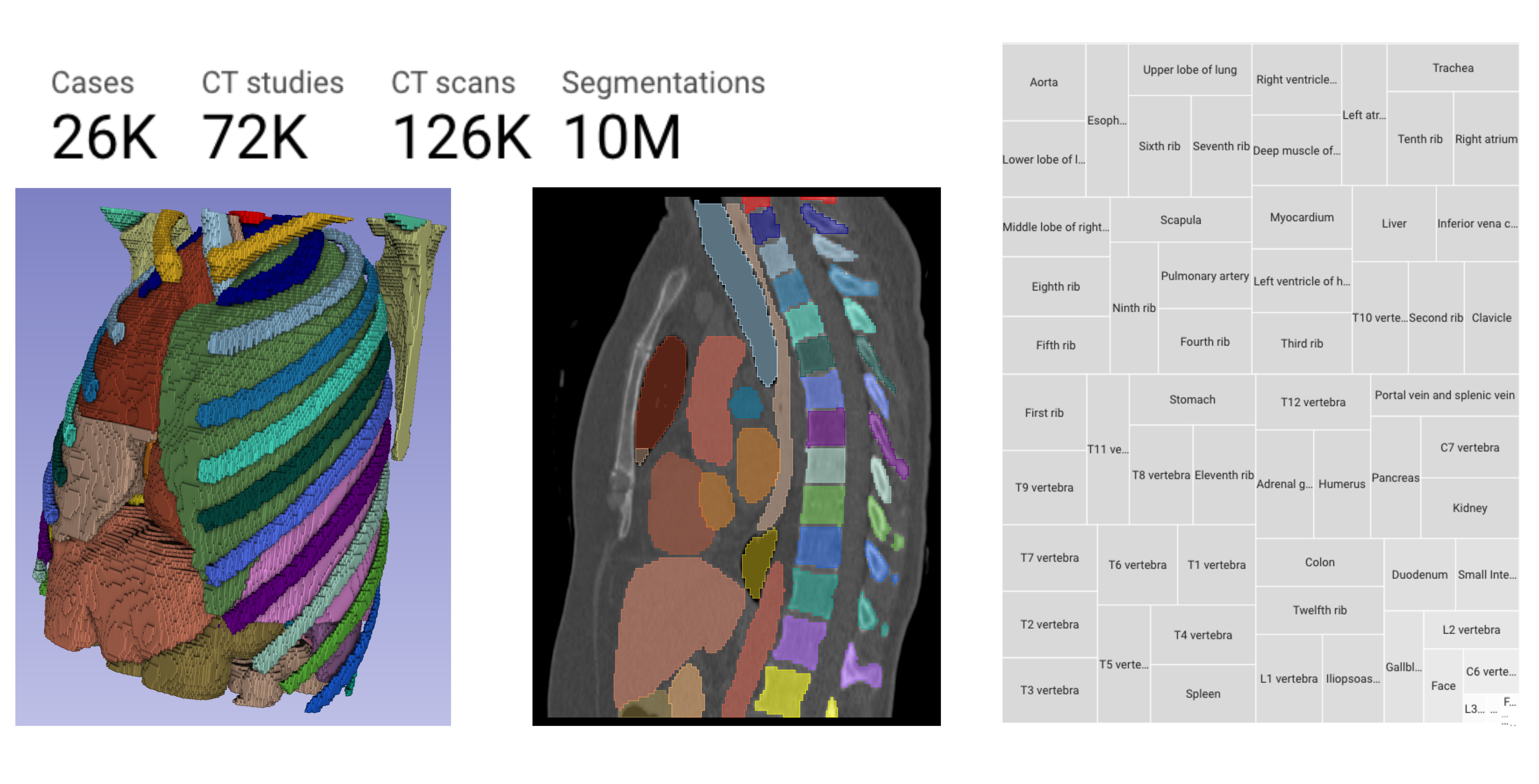
- The study successfully integrated this process within the UKSH MeDIC, resulting in the semantic enrichment of over 230,000 CT image series and over 8 million SNOMED CT annotations.
- The standardized representation using HL7 FHIR resources improves data discoverability and facilitates interoperability, providing a foundation for the FAIRness of medical imaging data.
Plain English Explanation
The paper describes how the researchers used AI to make medical imaging data, specifically CT scans, more useful and accessible. CT scans can contain a lot of valuable information, but it can be difficult to find and use that information, especially when working with large datasets from many different sources.
The researchers developed a process to automatically categorize and label the CT scans using a common medical terminology system called SNOMED CT. This makes it easier to search for and understand the information in the scans. They also standardized the data using a common medical data format called HL7 FHIR, which helps different computer systems communicate with each other more easily.
By doing this, the researchers were able to enhance over 230,000 CT image series with these semantic labels and annotations. This makes the data much more discoverable and shareable, which is important for advancing medical research and developing new AI-powered healthcare applications.
However, the researchers note that keeping up with the rapid growth of clinical datasets remains a challenge. Developing even more automated annotation methods will be key to ensuring continued progress in integrating and indexing large-scale medical imaging data.
Technical Explanation
The researchers developed an approach to semantically enhance clinical computed tomography (CT) image series to improve their findability, accessibility, interoperability, and reusability. Through an automated indexing process, the CT image series are annotated using the TotalSegmentator framework for segmentation and the resulting SNOMED CT terminology.
This standardized metadata representation using HL7 FHIR resources enables efficient data recognition and exchange between different research projects. The researchers successfully integrated this process within the UKSH MeDIC, leading to the semantic enrichment of over 230,000 CT image series and over 8 million SNOMED CT annotations.
The standardized FHIR-based representation improves the discoverability and interoperability of the medical imaging data, providing a foundation for the FAIRness of the datasets. However, the researchers note that developing automated annotation methods capable of keeping pace with the rapid growth of clinical datasets remains a challenge to ensure continued progress in large-scale integration and indexing of medical imaging for advanced healthcare AI applications.
Critical Analysis
The researchers have presented a robust approach to semantically enhancing clinical CT image data, which is a crucial step in making medical imaging more useful for advanced AI-powered healthcare applications. By standardizing the data representation and leveraging common medical terminology, the researchers have addressed key challenges around the findability, accessibility, interoperability, and reusability of these large and complex datasets.
However, the researchers acknowledge that the rapid growth of clinical datasets poses an ongoing challenge for developing automated annotation methods that can keep pace. Continued progress in this area will be essential to ensure the long-term sustainability and scalability of the proposed approach.
Additionally, the paper does not delve into the potential biases or limitations of the underlying AI models used for segmentation and annotation. As with any AI system, it is important to carefully evaluate the quality and representativeness of the training data to ensure the reliability and fairness of the resulting annotations.
Overall, the researchers have made an important contribution to the field of medical imaging informatics, but further work is needed to address the challenges of scaling these techniques to handle the ever-increasing volume and complexity of clinical data.
Conclusion
The integration of AI into medical imaging has led to significant advancements, but the reliability of these systems is heavily dependent on the quality and representativeness of the training data. This paper presents a promising approach to semantically enhancing clinical CT image series, which improves the findability, accessibility, interoperability, and reusability of these valuable datasets.
By leveraging standardized medical terminologies and data formats, the researchers have created a foundation for the FAIRness of medical imaging data, enabling more efficient data recognition and exchange between research projects. This is a crucial step in unlocking the full potential of AI-powered healthcare applications.
However, the rapid growth of clinical datasets remains a challenge, and the researchers acknowledge the need for continued development of automated annotation methods to ensure the scalability and sustainability of their approach. Addressing these ongoing challenges will be key to realizing the transformative impact of AI integration in the field of medical imaging.
This summary was produced with help from an AI and may contain inaccuracies - check out the links to read the original source documents!
Related Papers
🚀
0
Full-Scale Indexing and Semantic Annotation of CT Imaging: Boosting FAIRness
Hannes Ulrich, Robin Hendel, Santiago Pazmino, Bjorn Bergh, Bjorn Schreiweis
Background: The integration of artificial intelligence into medicine has led to significant advances, particularly in diagnostics and treatment planning. However, the reliability of AI models is highly dependent on the quality of the training data, especially in medical imaging, where varying patient data and evolving medical knowledge pose a challenge to the accuracy and generalizability of given datasets. Results: The proposed approach focuses on the integration and enhancement of clinical computed tomography (CT) image series for better findability, accessibility, interoperability, and reusability. Through an automated indexing process, CT image series are semantically enhanced using the TotalSegmentator framework for segmentation and resulting SNOMED CT annotations. The metadata is standardized with HL7 FHIR resources to enable efficient data recognition and data exchange between research projects. Conclusions: The study successfully integrates a robust process within the UKSH MeDIC, leading to the semantic enrichment of over 230,000 CT image series and over 8 million SNOMED CT annotations. The standardized representation using HL7 FHIR resources improves discoverability and facilitates interoperability, providing a foundation for the FAIRness of medical imaging data. However, developing automated annotation methods that can keep pace with growing clinical datasets remains a challenge to ensure continued progress in large-scale integration and indexing of medical imaging for advanced healthcare AI applications.
Read more6/24/2024

0
Coupling AI and Citizen Science in Creation of Enhanced Training Dataset for Medical Image Segmentation
Amir Syahmi, Xiangrong Lu, Yinxuan Li, Haoxuan Yao, Hanjun Jiang, Ishita Acharya, Shiyi Wang, Yang Nan, Xiaodan Xing, Guang Yang
Recent advancements in medical imaging and artificial intelligence (AI) have greatly enhanced diagnostic capabilities, but the development of effective deep learning (DL) models is still constrained by the lack of high-quality annotated datasets. The traditional manual annotation process by medical experts is time- and resource-intensive, limiting the scalability of these datasets. In this work, we introduce a robust and versatile framework that combines AI and crowdsourcing to improve both the quality and quantity of medical image datasets across different modalities. Our approach utilises a user-friendly online platform that enables a diverse group of crowd annotators to label medical images efficiently. By integrating the MedSAM segmentation AI with this platform, we accelerate the annotation process while maintaining expert-level quality through an algorithm that merges crowd-labelled images. Additionally, we employ pix2pixGAN, a generative AI model, to expand the training dataset with synthetic images that capture realistic morphological features. These methods are combined into a cohesive framework designed to produce an enhanced dataset, which can serve as a universal pre-processing pipeline to boost the training of any medical deep learning segmentation model. Our results demonstrate that this framework significantly improves model performance, especially when training data is limited.
Read more9/6/2024
0
Rule-based outlier detection of AI-generated anatomy segmentations
Deepa Krishnaswamy, Vamsi Krishna Thiriveedhi, Cosmin Ciausu, David Clunie, Steve Pieper, Ron Kikinis, Andrey Fedorov
There is a dire need for medical imaging datasets with accompanying annotations to perform downstream patient analysis. However, it is difficult to manually generate these annotations, due to the time-consuming nature, and the variability in clinical conventions. Artificial intelligence has been adopted in the field as a potential method to annotate these large datasets, however, a lack of expert annotations or ground truth can inhibit the adoption of these annotations. We recently made a dataset publicly available including annotations and extracted features of up to 104 organs for the National Lung Screening Trial using the TotalSegmentator method. However, the released dataset does not include expert-derived annotations or an assessment of the accuracy of the segmentations, limiting its usefulness. We propose the development of heuristics to assess the quality of the segmentations, providing methods to measure the consistency of the annotations and a comparison of results to the literature. We make our code and related materials publicly available at https://github.com/ImagingDataCommons/CloudSegmentatorResults and interactive tools at https://huggingface.co/spaces/ImagingDataCommons/CloudSegmentatorResults.
Read more6/21/2024

0
Charting the Path Forward: CT Image Quality Assessment -- An In-Depth Review
Siyi Xun, Qiaoyu Li, Xiaohong Liu, Guangtao Zhai, Mingxiang Wu, Tao Tan
Computed Tomography (CT) is a frequently utilized imaging technology that is employed in the clinical diagnosis of many disorders. However, clinical diagnosis, data storage, and management are posed huge challenges by a huge volume of non-homogeneous CT data in terms of imaging quality. As a result, the quality assessment of CT images is a crucial problem that demands consideration. The history, advancements in research, and current developments in CT image quality assessment (IQA) are examined in this paper. In this review, we collected and researched more than 500 CT-IQA publications published before August 2023. And we provide the visualization analysis of keywords and co-citations in the knowledge graph of these papers. Prospects and obstacles for the continued development of CT-IQA are also covered. At present, significant research branches in the CT-IQA domain include Phantom study, Artificial intelligence deep-learning reconstruction algorithm, Dose reduction opportunity, and Virtual monoenergetic reconstruction. Artificial intelligence (AI)-based CT-IQA also becomes a trend. It increases the accuracy of the CT scanning apparatus, amplifies the impact of the CT system reconstruction algorithm, and creates an effective algorithm for post-processing CT images. AI-based medical IQA offers excellent application opportunities in clinical work. AI can provide uniform quality assessment criteria and more comprehensive guidance amongst various healthcare facilities, and encourage them to identify one another's images. It will help lower the number of unnecessary tests and associated costs, and enhance the quality of medical imaging and assessment efficiency.
Read more5/2/2024