Charting the Path Forward: CT Image Quality Assessment -- An In-Depth Review

0

Sign in to get full access
Overview
- This paper provides an in-depth review of CT image quality assessment, exploring the current state of the field and charting a path forward.
- The review covers various aspects of CT image quality assessment, including the background, existing methods, and emerging trends.
- It highlights the importance of accurate CT image quality assessment in medical diagnosis and treatment planning, and the need for continued research and development in this area.
Plain English Explanation
Computed tomography (CT) scans are an essential medical imaging technique that allow doctors to see detailed images of the inside of the body. However, the quality of these images can vary, and it's important to be able to accurately assess and measure this quality. This paper reviews the current state of research on assessing the quality of CT images, and suggests ways to improve and advance this field.
The paper starts by providing background information on CT imaging and the importance of image quality assessment. It then goes on to discuss the various methods and approaches that have been developed to evaluate the quality of CT images. These include evaluating the image quality of CT scans using simple generative representation, using large multi-modality models to assist in AI-generated CT images, and leveraging perceptual quality assessment databases like the PKU-AIGIQA 4K dataset.
The paper then discusses emerging trends and future directions in CT image quality assessment, including the potential for autonomous quality hallucination assessment for virtual tissue staining and the development of more advanced image quality assessment models using compressive sampling.
Overall, this paper provides a comprehensive overview of the current state of CT image quality assessment and highlights the importance of continued research and innovation in this field to improve medical diagnosis and treatment.
Technical Explanation
The paper begins by providing background on the importance of CT image quality assessment in medical diagnosis and treatment planning. It outlines the various factors that can affect CT image quality, such as radiation dose, patient movement, and imaging hardware and software.
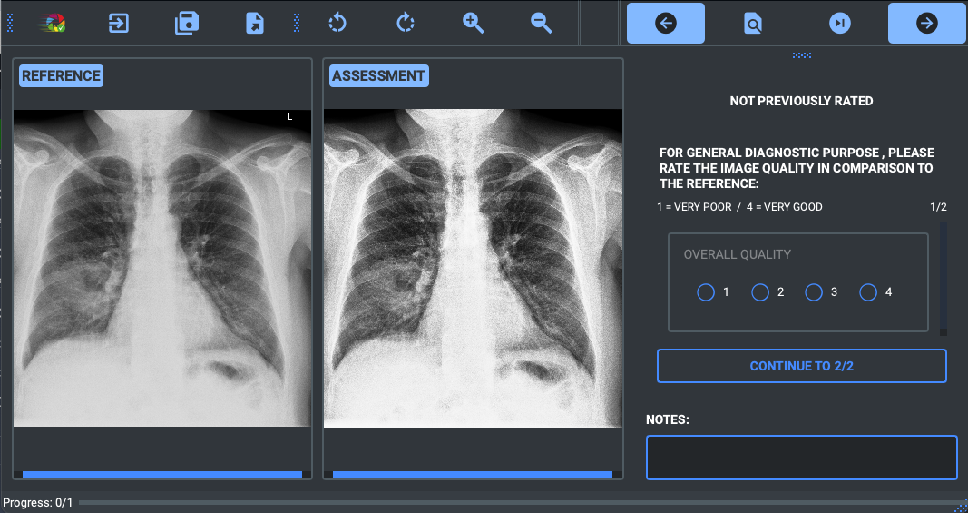
The review then delves into the various methods and approaches that have been developed for assessing CT image quality. This includes both subjective and objective assessment techniques. Subjective methods rely on human evaluators to rate image quality, while objective methods use computational algorithms to measure specific image quality metrics.
The paper discusses several state-of-the-art approaches, such as using simple generative representation to assess image quality, leveraging large multi-modality models to assist in AI-generated CT images, and utilizing perceptual quality assessment databases like the PKU-AIGIQA 4K dataset. It also highlights emerging trends, including autonomous quality hallucination assessment for virtual tissue staining and the development of advanced image quality assessment models using compressive sampling.
The paper provides a comprehensive and in-depth review of the current state of CT image quality assessment, identifying key challenges, advancements, and future research directions in this critical field.
Critical Analysis
The paper provides a thorough and well-structured review of CT image quality assessment, covering a broad range of existing methods and emerging trends. However, it is important to note that the field is rapidly evolving, and some of the information and techniques discussed may become outdated quickly.
One potential limitation of the review is that it does not dive deeply into the specific technical details and implementation challenges of the various approaches discussed. While this is understandable given the broad scope of the paper, it may leave some readers wanting more in-depth technical information.
Additionally, the paper does not extensively discuss the potential biases and limitations of the various assessment methods, such as the subjectivity inherent in human-based evaluations or the potential for algorithmic biases in computational approaches. Addressing these issues could have strengthened the critical analysis and provided readers with a more nuanced understanding of the field.
Despite these minor limitations, the paper provides a valuable and comprehensive overview of the current state of CT image quality assessment, and serves as a useful resource for researchers and practitioners in this field. Encouraging readers to think critically about the research and form their own opinions is an important aspect of this review.
Conclusion
This paper offers a comprehensive and in-depth review of the current state of CT image quality assessment, providing a strong foundation for understanding the key challenges, advancements, and future directions in this critical field. By covering a wide range of existing methods and emerging trends, the review highlights the importance of accurate and reliable CT image quality assessment in medical diagnosis and treatment planning.
The paper's discussion of state-of-the-art approaches, such as the use of simple generative representation, large multi-modality models, and perceptual quality assessment databases, as well as emerging trends like autonomous quality hallucination assessment and advanced image quality assessment models using compressive sampling, provides valuable insights for researchers and practitioners looking to advance the field.
While the paper acknowledges some potential limitations and areas for further research, its comprehensive coverage and critical analysis make it a valuable resource for understanding the current state of CT image quality assessment and charting a path forward for continued progress in this important area of medical imaging.
This summary was produced with help from an AI and may contain inaccuracies - check out the links to read the original source documents!
Related Papers
0
Charting the Path Forward: CT Image Quality Assessment -- An In-Depth Review
Siyi Xun, Qiaoyu Li, Xiaohong Liu, Guangtao Zhai, Mingxiang Wu, Tao Tan
Computed Tomography (CT) is a frequently utilized imaging technology that is employed in the clinical diagnosis of many disorders. However, clinical diagnosis, data storage, and management are posed huge challenges by a huge volume of non-homogeneous CT data in terms of imaging quality. As a result, the quality assessment of CT images is a crucial problem that demands consideration. The history, advancements in research, and current developments in CT image quality assessment (IQA) are examined in this paper. In this review, we collected and researched more than 500 CT-IQA publications published before August 2023. And we provide the visualization analysis of keywords and co-citations in the knowledge graph of these papers. Prospects and obstacles for the continued development of CT-IQA are also covered. At present, significant research branches in the CT-IQA domain include Phantom study, Artificial intelligence deep-learning reconstruction algorithm, Dose reduction opportunity, and Virtual monoenergetic reconstruction. Artificial intelligence (AI)-based CT-IQA also becomes a trend. It increases the accuracy of the CT scanning apparatus, amplifies the impact of the CT system reconstruction algorithm, and creates an effective algorithm for post-processing CT images. AI-based medical IQA offers excellent application opportunities in clinical work. AI can provide uniform quality assessment criteria and more comprehensive guidance amongst various healthcare facilities, and encourage them to identify one another's images. It will help lower the number of unnecessary tests and associated costs, and enhance the quality of medical imaging and assessment efficiency.
Read more5/2/2024

0
A study of why we need to reassess full reference image quality assessment with medical images
Anna Breger, Ander Biguri, Malena Sabat'e Landman, Ian Selby, Nicole Amberg, Elisabeth Brunner, Janek Grohl, Sepideh Hatamikia, Clemens Karner, Lipeng Ning, Soren Dittmer, Michael Roberts, AIX-COVNET Collaboration, Carola-Bibiane Schonlieb
Image quality assessment (IQA) is not just indispensable in clinical practice to ensure high standards, but also in the development stage of novel algorithms that operate on medical images with reference data. This paper provides a structured and comprehensive collection of examples where the two most common full reference (FR) image quality measures prove to be unsuitable for the assessment of novel algorithms using different kinds of medical images, including real-world MRI, CT, OCT, X-Ray, digital pathology and photoacoustic imaging data. In particular, the FR-IQA measures PSNR and SSIM are known and tested for working successfully in many natural imaging tasks, but discrepancies in medical scenarios have been noted in the literature. Inconsistencies arising in medical images are not surprising, as they have very different properties than natural images which have not been targeted nor tested in the development of the mentioned measures, and therefore might imply wrong judgement of novel methods for medical images. Therefore, improvement is urgently needed in particular in this era of AI to increase explainability, reproducibility and generalizability in machine learning for medical imaging and beyond. On top of the pitfalls we will provide ideas for future research as well as suggesting guidelines for the usage of FR-IQA measures applied to medical images.
Read more5/30/2024

0
Rethinking Medical Anomaly Detection in Brain MRI: An Image Quality Assessment Perspective
Zixuan Pan, Jun Xia, Zheyu Yan, Guoyue Xu, Yawen Wu, Zhenge Jia, Jianxu Chen, Yiyu Shi
Reconstruction-based methods, particularly those leveraging autoencoders, have been widely adopted to perform anomaly detection in brain MRI. While most existing works try to improve detection accuracy by proposing new model structures or algorithms, we tackle the problem through image quality assessment, an underexplored perspective in the field. We propose a fusion quality loss function that combines Structural Similarity Index Measure loss with l1 loss, offering a more comprehensive evaluation of reconstruction quality. Additionally, we introduce a data pre-processing strategy that enhances the average intensity ratio (AIR) between normal and abnormal regions, further improving the distinction of anomalies. By fusing the aforementioned two methods, we devise the image quality assessment (IQA) approach. The proposed IQA approach achieves significant improvements (>10%) in terms of Dice coefficient (DICE) and Area Under the Precision-Recall Curve (AUPRC) on the BraTS21 (T2, FLAIR) and MSULB datasets when compared with state-of-the-art methods. These results highlight the importance of invoking the comprehensive image quality assessment in medical anomaly detection and provide a new perspective for future research in this field.
Read more8/16/2024
0
A study on the adequacy of common IQA measures for medical images
Anna Breger, Clemens Karner, Ian Selby, Janek Grohl, Soren Dittmer, Edward Lilley, Judith Babar, Jake Beckford, Thomas R Else, Timothy J Sadler, Shahab Shahipasand, Arthikkaa Thavakumar, Michael Roberts, Carola-Bibiane Schonlieb
Image quality assessment (IQA) is standard practice in the development stage of novel machine learning algorithms that operate on images. The most commonly used IQA measures have been developed and tested for natural images, but not in the medical setting. Reported inconsistencies arising in medical images are not surprising, as they have different properties than natural images. In this study, we test the applicability of common IQA measures for medical image data by comparing their assessment to manually rated chest X-ray (5 experts) and photoacoustic image data (2 experts). Moreover, we include supplementary studies on grayscale natural images and accelerated brain MRI data. The results of all experiments show a similar outcome in line with previous findings for medical imaging: PSNR and SSIM in the default setting are in the lower range of the result list and HaarPSI outperforms the other tested measures in the overall performance. Also among the top performers in our medical experiments are the full reference measures FSIM, GMSD, LPIPS and MS-SSIM. Generally, the results on natural images yield considerably higher correlations, suggesting that the additional employment of tailored IQA measures for medical imaging algorithms is needed.
Read more8/21/2024