Gallbladder Cancer Detection in Ultrasound Images based on YOLO and Faster R-CNN

0
🔎
Sign in to get full access
Overview
- This paper explores the use of object detection algorithms, specifically YOLO and Faster R-CNN, for the identification of gallbladder regions in ultrasound images to enhance gallbladder cancer classification.
- The study proposes a fusion method that combines the strengths of both techniques to improve the accuracy of bounding box detection compared to using the individual algorithms.
- The proposed method demonstrated superior classification performance, with an accuracy of 92.62%, outperforming the individual use of Faster R-CNN and YOLOv8, which yielded accuracies of 90.16% and 82.79%, respectively.
Plain English Explanation
Medical image analysis is a crucial application of artificial intelligence (AI) for disease diagnosis. One key step in this process is the identification of specific regions within the images that are important for analysis, known as regions of interest. This task can be automated using object detection algorithms, such as YOLO and Faster R-CNN, which are well-known for their capabilities.
In this study, the researchers aimed to explore the advantages of both YOLO and Faster R-CNN techniques to select more accurate bounding boxes (the rectangular areas that identify the objects of interest) for detecting the gallbladder in ultrasound images. This is an important step for improving the accuracy of gallbladder cancer classification, which is a type of cancer detection using AI.
The researchers proposed a new method that combines the strengths of both YOLO and Faster R-CNN, creating a "fusion" approach. This fusion method was found to be more accurate than using either YOLO or Faster R-CNN alone, achieving an accuracy of 92.62% in gallbladder cancer classification, compared to 90.16% for Faster R-CNN and 82.79% for YOLOv8 (the latest version of YOLO).
Technical Explanation
The study aimed to explore the advantages of the YOLO and Faster R-CNN object detection algorithms for accurately identifying the gallbladder region in ultrasound images, which is a crucial step for classifying gallbladder cancer.
The researchers implemented both YOLO and Faster R-CNN models and evaluated their performance on a dataset of ultrasound images. They then proposed a fusion method that combines the strengths of both techniques to improve the accuracy of bounding box detection for the gallbladder.
The fusion method involved using the bounding boxes generated by both YOLO and Faster R-CNN, and then applying a heuristic algorithm to select the most accurate bounding box. This approach aimed to leverage the benefits of each algorithm, such as YOLO's speed and Faster R-CNN's precision, to achieve better overall performance.
The results showed that the proposed fusion method outperformed the individual use of YOLO and Faster R-CNN, achieving an accuracy of 92.62% in gallbladder cancer classification, compared to 90.16% for Faster R-CNN and 82.79% for YOLOv8.
Critical Analysis
The study presents a promising approach for improving gallbladder cancer detection by leveraging the strengths of two popular object detection algorithms. However, there are a few potential limitations and areas for further research:
-
The study was conducted on a specific dataset of ultrasound images, and it's unclear how the fusion method would perform on a more diverse set of medical images or in real-world clinical settings. Further research is needed to assess the generalizability of the approach.
-
The study does not provide detailed information about the dataset, such as the number of samples, the distribution of benign and malignant cases, and the characteristics of the images. This makes it difficult to fully evaluate the robustness and reliability of the results.
-
While the fusion method demonstrated improved accuracy compared to the individual algorithms, the study could have explored other fusion techniques or ensembling strategies to further enhance the performance.
-
The paper does not discuss the computational complexity or inference time of the proposed fusion method, which are important factors to consider for real-time medical applications.
Overall, the study presents a valuable contribution to the field of medical image analysis, but additional research and validation would be beneficial to fully understand the strengths, limitations, and practical implications of the proposed fusion approach.
Conclusion
This study explored the use of object detection algorithms, specifically YOLO and Faster R-CNN, for the identification of gallbladder regions in ultrasound images to enhance gallbladder cancer classification. The researchers proposed a fusion method that combines the strengths of both techniques, resulting in improved bounding box detection and a classification accuracy of 92.62%.
The findings of this study demonstrate the potential of leveraging multiple object detection algorithms to enhance medical image analysis tasks, such as cancer detection. The fusion approach presented in this paper could have broader applications in various medical imaging domains, potentially leading to more accurate and reliable disease diagnosis and treatment planning.
This summary was produced with help from an AI and may contain inaccuracies - check out the links to read the original source documents!
Related Papers
🔎
0
Gallbladder Cancer Detection in Ultrasound Images based on YOLO and Faster R-CNN
Sara Dadjouy, Hedieh Sajedi
Medical image analysis is a significant application of artificial intelligence for disease diagnosis. A crucial step in this process is the identification of regions of interest within the images. This task can be automated using object detection algorithms. YOLO and Faster R-CNN are renowned for such algorithms, each with its own strengths and weaknesses. This study aims to explore the advantages of both techniques to select more accurate bounding boxes for gallbladder detection from ultrasound images, thereby enhancing gallbladder cancer classification. A fusion method that leverages the benefits of both techniques is presented in this study. The proposed method demonstrated superior classification performance, with an accuracy of 92.62%, compared to the individual use of Faster R-CNN and YOLOv8, which yielded accuracies of 90.16% and 82.79%, respectively.
Read more4/24/2024
🖼️
0
MedYOLO: A Medical Image Object Detection Framework
Joseph Sobek, Jose R. Medina Inojosa, Betsy J. Medina Inojosa, S. M. Rassoulinejad-Mousavi, Gian Marco Conte, Francisco Lopez-Jimenez, Bradley J. Erickson
Artificial intelligence-enhanced identification of organs, lesions, and other structures in medical imaging is typically done using convolutional neural networks (CNNs) designed to make voxel-accurate segmentations of the region of interest. However, the labels required to train these CNNs are time-consuming to generate and require attention from subject matter experts to ensure quality. For tasks where voxel-level precision is not required, object detection models offer a viable alternative that can reduce annotation effort. Despite this potential application, there are few options for general purpose object detection frameworks available for 3-D medical imaging. We report on MedYOLO, a 3-D object detection framework using the one-shot detection method of the YOLO family of models and designed for use with medical imaging. We tested this model on four different datasets: BRaTS, LIDC, an abdominal organ Computed Tomography (CT) dataset, and an ECG-gated heart CT dataset. We found our models achieve high performance on commonly present medium and large-sized structures such as the heart, liver, and pancreas even without hyperparameter tuning. However, the models struggle with very small or rarely present structures.
Read more6/10/2024

0
LSM-YOLO: A Compact and Effective ROI Detector for Medical Detection
Zhongwen Yu, Qiu Guan, Jianmin Yang, Zhiqiang Yang, Qianwei Zhou, Yang Chen, Feng Chen
In existing medical Region of Interest (ROI) detection, there lacks an algorithm that can simultaneously satisfy both real-time performance and accuracy, not meeting the growing demand for automatic detection in medicine. Although the basic YOLO framework ensures real-time detection due to its fast speed, it still faces challenges in maintaining precision concurrently. To alleviate the above problems, we propose a novel model named Lightweight Shunt Matching-YOLO (LSM-YOLO), with Lightweight Adaptive Extraction (LAE) and Multipath Shunt Feature Matching (MSFM). Firstly, by using LAE to refine feature extraction, the model can obtain more contextual information and high-resolution details from multiscale feature maps, thereby extracting detailed features of ROI in medical images while reducing the influence of noise. Secondly, MSFM is utilized to further refine the fusion of high-level semantic features and low-level visual features, enabling better fusion between ROI features and neighboring features, thereby improving the detection rate for better diagnostic assistance. Experimental results demonstrate that LSM-YOLO achieves 48.6% AP on a private dataset of pancreatic tumors, 65.1% AP on the BCCD blood cell detection public dataset, and 73.0% AP on the Br35h brain tumor detection public dataset. Our model achieves state-of-the-art performance with minimal parameter cost on the above three datasets. The source codes are at: https://github.com/VincentYuuuuuu/LSM-YOLO.
Read more8/27/2024
0
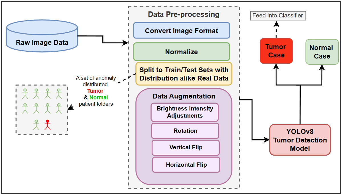
Realism in Action: Anomaly-Aware Diagnosis of Brain Tumors from Medical Images Using YOLOv8 and DeiT
Seyed Mohammad Hossein Hashemi, Leila Safari, Amirhossein Dadashzadeh Taromi
In the field of medical sciences, reliable detection and classification of brain tumors from images remains a formidable challenge due to the rarity of tumors within the population of patients. Therefore, the ability to detect tumors in anomaly scenarios is paramount for ensuring timely interventions and improved patient outcomes. This study addresses the issue by leveraging deep learning (DL) techniques to detect and classify brain tumors in challenging situations. The curated data set from the National Brain Mapping Lab (NBML) comprises 81 patients, including 30 Tumor cases and 51 Normal cases. The detection and classification pipelines are separated into two consecutive tasks. The detection phase involved comprehensive data analysis and pre-processing to modify the number of image samples and the number of patients of each class to anomaly distribution (9 Normal per 1 Tumor) to comply with real world scenarios. Next, in addition to common evaluation metrics for the testing, we employed a novel performance evaluation method called Patient to Patient (PTP), focusing on the realistic evaluation of the model. In the detection phase, we fine-tuned a YOLOv8n detection model to detect the tumor region. Subsequent testing and evaluation yielded competitive performance both in Common Evaluation Metrics and PTP metrics. Furthermore, using the Data Efficient Image Transformer (DeiT) module, we distilled a Vision Transformer (ViT) model from a fine-tuned ResNet152 as a teacher in the classification phase. This approach demonstrates promising strides in reliable tumor detection and classification, offering potential advancements in tumor diagnosis for real-world medical imaging scenarios.
Read more9/26/2024