Generative artificial intelligence in ophthalmology: multimodal retinal images for the diagnosis of Alzheimer's disease with convolutional neural networks

0
Sign in to get full access
Overview
- This research paper explores the use of generative artificial intelligence (AI) in ophthalmology to diagnose Alzheimer's disease using multimodal retinal images and convolutional neural networks.
- The study aims to develop a non-invasive, low-cost method for early detection of Alzheimer's disease by analyzing changes in the retina, which is connected to the brain.
- The researchers employed a deep learning approach, specifically convolutional neural networks, to classify Alzheimer's disease based on different types of retinal images, including color fundus, optical coherence tomography (OCT), and autofluorescence.
Plain English Explanation
The researchers in this study wanted to find a way to diagnose Alzheimer's disease, a type of dementia, using images of the eye. Alzheimer's disease is a serious condition that affects the brain, and it can be difficult to diagnose early on. The researchers thought that changes in the retina, the light-sensitive layer at the back of the eye, might be a sign of Alzheimer's disease, since the retina is connected to the brain.
To test this idea, the researchers used a type of artificial intelligence called a convolutional neural network. This is a type of machine learning algorithm that can analyze and classify images. The researchers trained the neural network to look for patterns in different types of retinal images, including color photos of the retina, images taken with a special camera that measures the thickness of the retina (called optical coherence tomography or OCT), and images that show the glow of certain molecules in the retina (called autofluorescence).
By analyzing these retinal images, the researchers were able to develop a non-invasive and relatively low-cost way to detect Alzheimer's disease. This is important because current methods for diagnosing Alzheimer's disease can be expensive and involve procedures that are uncomfortable for the patient, such as spinal taps or brain scans. The researchers hope that their approach using AI and retinal images could lead to earlier detection of Alzheimer's disease, which could help patients receive treatment and support sooner.
Technical Explanation
The researchers in this study used a deep learning approach, specifically convolutional neural networks, to classify Alzheimer's disease based on different types of retinal images, including color fundus, optical coherence tomography (OCT), and autofluorescence.
The study design involved training and evaluating the deep learning models using a dataset of retinal images from individuals with and without Alzheimer's disease. The researchers pre-processed the images, augmented the data, and then trained the convolutional neural networks to learn features that could distinguish between the two groups.
The key architectural elements of the models included the use of multiple convolutional layers to extract relevant visual features, followed by pooling layers to reduce the dimensionality of the data, and finally, fully connected layers to perform the classification task. The researchers also explored the use of transfer learning, where pre-trained models on large-scale image datasets were fine-tuned on the retinal image data to improve performance.
The results of the study demonstrated that the deep learning models were able to achieve high accuracy, sensitivity, and specificity in diagnosing Alzheimer's disease using the multimodal retinal images. The researchers found that the combined use of different retinal imaging modalities, such as color fundus, OCT, and autofluorescence, led to improved performance compared to using a single modality.
The insights gained from this research suggest that generative AI techniques in ophthalmology, specifically the use of convolutional neural networks on multimodal retinal images, can be a promising approach for the non-invasive, early detection of Alzheimer's disease. This could have significant implications for improving patient outcomes and reducing the burden of this devastating neurodegenerative disorder.
Critical Analysis
The researchers acknowledge several limitations and areas for future research in their paper. One key limitation is the relatively small sample size of the dataset, which may affect the generalizability of the results. Additionally, the study was conducted on a specific population, and further validation is needed to ensure the approach can be applied effectively across diverse demographic groups.
Another potential issue is the interpretability of the deep learning models. While the models demonstrated high accuracy, it can be challenging to understand the specific features or biomarkers that the models are using to make their diagnoses. This lack of interpretability can be a concern, as it can make it difficult to validate the models' decision-making process and understand the underlying mechanisms linking retinal changes to Alzheimer's disease.
Future research could focus on developing more interpretable deep learning models, perhaps using techniques like Grad-CAM or exploring multi-view, multi-modal approaches, to provide better insights into the relationship between retinal features and Alzheimer's disease. Additionally, longitudinal studies and larger, more diverse datasets would be valuable to further validate the generalizability and clinical utility of this approach.
Conclusion
This research demonstrates the potential of generative AI techniques, specifically convolutional neural networks, in the field of ophthalmology for the early detection of Alzheimer's disease. By leveraging multimodal retinal images, the researchers have developed a non-invasive, low-cost method that could enable earlier diagnosis and intervention for this debilitating neurodegenerative disorder.
While the results are promising, further research is needed to address the limitations and improve the interpretability of the deep learning models. Nonetheless, this study represents an important step forward in the use of AI for Alzheimer's disease classification and multimodal neuroimaging approaches that could ultimately lead to better outcomes for patients and their families.
This summary was produced with help from an AI and may contain inaccuracies - check out the links to read the original source documents!
Related Papers
0
Generative artificial intelligence in ophthalmology: multimodal retinal images for the diagnosis of Alzheimer's disease with convolutional neural networks
I. R. Slootweg, M. Thach, K. R. Curro-Tafili, F. D. Verbraak, F. H. Bouwman, Y. A. L. Pijnenburg, J. F. Boer, J. H. P. de Kwisthout, L. Bagheriye, P. J. Gonz'alez
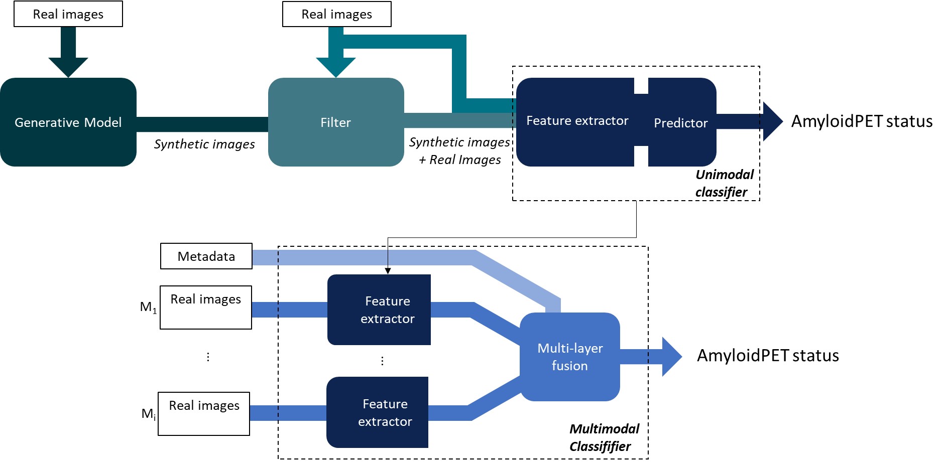
Background/Aim. This study aims to predict Amyloid Positron Emission Tomography (AmyloidPET) status with multimodal retinal imaging and convolutional neural networks (CNNs) and to improve the performance through pretraining with synthetic data. Methods. Fundus autofluorescence, optical coherence tomography (OCT), and OCT angiography images from 328 eyes of 59 AmyloidPET positive subjects and 108 AmyloidPET negative subjects were used for classification. Denoising Diffusion Probabilistic Models (DDPMs) were trained to generate synthetic images and unimodal CNNs were pretrained on synthetic data and finetuned on real data or trained solely on real data. Multimodal classifiers were developed to combine predictions of the four unimodal CNNs with patient metadata. Class activation maps of the unimodal classifiers provided insight into the network's attention to inputs. Results. DDPMs generated diverse, realistic images without memorization. Pretraining unimodal CNNs with synthetic data improved AUPR at most from 0.350 to 0.579. Integration of metadata in multimodal CNNs improved AUPR from 0.486 to 0.634, which was the best overall best classifier. Class activation maps highlighted relevant retinal regions which correlated with AD. Conclusion. Our method for generating and leveraging synthetic data has the potential to improve AmyloidPET prediction from multimodal retinal imaging. A DDPM can generate realistic and unique multimodal synthetic retinal images. Our best performing unimodal and multimodal classifiers were not pretrained on synthetic data, however pretraining with synthetic data slightly improved classification performance for two out of the four modalities.
Read more6/27/2024

0
Enhancing Eye Disease Diagnosis with Deep Learning and Synthetic Data Augmentation
Saideep Kilaru, Kothamasu Jayachandra, Tanishka Yagneshwar, Suchi Kumari
In recent years, the focus is on improving the diagnosis of diabetic retinopathy (DR) using machine learning and deep learning technologies. Researchers have explored various approaches, including the use of high-definition medical imaging, AI-driven algorithms such as convolutional neural networks (CNNs) and generative adversarial networks (GANs). Among all the available tools, CNNs have emerged as a preferred tool due to their superior classification accuracy and efficiency. Although the accuracy of CNNs is comparatively better but it can be improved by introducing some hybrid models by combining various machine learning and deep learning models. Therefore, in this paper, an ensemble learning technique is proposed for early detection and management of DR with higher accuracy. The proposed model is tested on the APTOS dataset and it is showing supremacy on the validation accuracy ($99%)$ in comparison to the previous models. Hence, the model can be helpful for early detection and treatment of the DR, thereby enhancing the overall quality of care for affected individuals.
Read more7/26/2024

0
Self-Supervised Pretext Tasks for Alzheimer's Disease Classification using 3D Convolutional Neural Networks on Large-Scale Synthetic Neuroimaging Dataset
Chen Zheng
Structural magnetic resonance imaging (MRI) studies have shown that Alzheimer's Disease (AD) induces both localised and widespread neural degenerative changes throughout the brain. However, the absence of segmentation that highlights brain degenerative changes presents unique challenges for training CNN-based classifiers in a supervised fashion. In this work, we evaluated several unsupervised methods to train a feature extractor for downstream AD vs. CN classification. Using the 3D T1-weighted MRI data of cognitive normal (CN) subjects from the synthetic neuroimaging LDM100K dataset, lightweight 3D CNN-based models are trained for brain age prediction, brain image rotation classification, brain image reconstruction and a multi-head task combining all three tasks into one. Feature extractors trained on the LDM100K synthetic dataset achieved similar performance compared to the same model using real-world data. This supports the feasibility of utilising large-scale synthetic data for pretext task training. All the training and testing splits are performed on the subject-level to prevent data leakage issues. Alongside the simple preprocessing steps, the random cropping data augmentation technique shows consistent improvement across all experiments.
Read more6/21/2024

0
Multi-OCT-SelfNet: Integrating Self-Supervised Learning with Multi-Source Data Fusion for Enhanced Multi-Class Retinal Disease Classification
Fatema-E- Jannat, Sina Gholami, Jennifer I. Lim, Theodore Leng, Minhaj Nur Alam, Hamed Tabkhi
In the medical domain, acquiring large datasets poses significant challenges due to privacy concerns. Nonetheless, the development of a robust deep-learning model for retinal disease diagnosis necessitates a substantial dataset for training. The capacity to generalize effectively on smaller datasets remains a persistent challenge. The scarcity of data presents a significant barrier to the practical implementation of scalable medical AI solutions. To address this issue, we've combined a wide range of data sources to improve performance and generalization to new data by giving it a deeper understanding of the data representation from multi-modal datasets and developed a self-supervised framework based on large language models (LLMs), SwinV2 to gain a deeper understanding of multi-modal dataset representations, enhancing the model's ability to extrapolate to new data for the detection of eye diseases using optical coherence tomography (OCT) images. We adopt a two-phase training methodology, self-supervised pre-training, and fine-tuning on a downstream supervised classifier. An ablation study conducted across three datasets employing various encoder backbones, without data fusion, with low data availability setting, and without self-supervised pre-training scenarios, highlights the robustness of our method. Our findings demonstrate consistent performance across these diverse conditions, showcasing superior generalization capabilities compared to the baseline model, ResNet-50.
Read more9/18/2024