Hyperparameter-Free Medical Image Synthesis for Sharing Data and Improving Site-Specific Segmentation

0
Sign in to get full access
Overview
- This paper presents a novel method for generating realistic synthetic medical images that can be used to improve the performance of medical image segmentation models, particularly in cases where data is limited or unevenly distributed across different clinical sites.
- The method is "hyperparameter-free," meaning it does not require extensive tuning of model parameters, making it more accessible and easier to apply in real-world settings.
- The authors demonstrate the effectiveness of their approach on several medical imaging tasks, including brain MRI and chest X-ray segmentation, showing that the synthetic data can significantly boost the performance of segmentation models.
Plain English Explanation
The paper discusses a new way to create fake medical images that look very realistic. These synthetic images can be used to help train computer models that are used to analyze real medical scans, like MRI or X-ray images.
One of the challenges in medical imaging is that there often isn't enough real data available to train these models, especially if the data comes from different hospitals or clinics. The synthetic images created by the method described in this paper can be used to supplement the real data, helping the models learn more effectively.
Importantly, this new method is "hyperparameter-free," which means it doesn't require a lot of manual tuning and adjusting of the model's parameters to get good results. This makes it easier for doctors and researchers to use in practice, without needing a lot of specialized machine learning expertise.
The authors tested their approach on several medical imaging tasks, such as segmenting brain structures in MRI scans and detecting abnormalities in chest X-rays. They found that the synthetic data helped improve the performance of the segmentation models, especially in cases where the real data was limited or came from different clinical sites.
Technical Explanation
The key innovation of this paper is a hyperparameter-free medical image synthesis method that can generate realistic synthetic images to supplement limited real-world training data. The authors use a generative adversarial network (GAN) architecture, with a novel training procedure that does not require extensive tuning of GAN hyperparameters.
The method works by training the GAN on a mix of real and synthetic images, with the goal of making the synthetic images indistinguishable from the real ones. This "self-supervised" training approach allows the model to learn the underlying structure and characteristics of the medical images without relying on manual annotations or segmentation labels.
To evaluate the effectiveness of the synthetic data, the authors integrated it into the training of several segmentation models across different medical imaging modalities, including brain MRI and chest X-ray. They demonstrated that the synthetic data can significantly improve segmentation performance, especially in cases where the real-world training data is limited or unevenly distributed across clinical sites.
Critical Analysis
The authors acknowledge several limitations of their approach. First, while the method is "hyperparameter-free" in the sense that it does not require extensive tuning of GAN-specific hyperparameters, there are still other hyperparameters (e.g., optimizer settings, network architecture) that may need to be adjusted for optimal performance on specific tasks.
Additionally, the authors note that their approach assumes the availability of a diverse set of real medical images for the GAN to learn from. In cases where the real-world data is heavily biased or skewed, the synthetic images may inherit these same biases, which could negatively impact downstream segmentation models.
Furthermore, the authors do not provide a detailed analysis of the potential risks or ethical considerations around the use of synthetic medical data, such as concerns about privacy, data provenance, or the potential for misuse. These are important factors that should be carefully considered before deploying such systems in real-world clinical settings.
Despite these limitations, the work presented in this paper represents an important contribution to the field of medical image synthesis and has the potential to significantly improve the performance and robustness of medical image analysis models, especially in data-scarce scenarios.
Conclusion
This paper introduces a novel, hyperparameter-free method for generating synthetic medical images that can be used to improve the performance of medical image segmentation models. The authors demonstrate the effectiveness of their approach on several medical imaging tasks, showing that the synthetic data can help boost the accuracy of segmentation models, particularly in cases where the real-world training data is limited or unevenly distributed across different clinical sites.
While the method has some limitations, it represents an important step forward in the field of medical image synthesis and has the potential to significantly impact the development and deployment of more robust and reliable medical imaging AI systems.
This summary was produced with help from an AI and may contain inaccuracies - check out the links to read the original source documents!
Related Papers
0
Hyperparameter-Free Medical Image Synthesis for Sharing Data and Improving Site-Specific Segmentation
Alexander Chebykin, Peter A. N. Bosman, Tanja Alderliesten
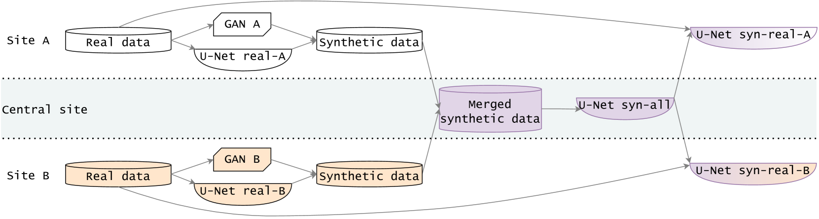
Sharing synthetic medical images is a promising alternative to sharing real images that can improve patient privacy and data security. To get good results, existing methods for medical image synthesis must be manually adjusted when they are applied to unseen data. To remove this manual burden, we introduce a Hyperparameter-Free distributed learning method for automatic medical image Synthesis, Sharing, and Segmentation called HyFree-S3. For three diverse segmentation settings (pelvic MRIs, lung X-rays, polyp photos), the use of HyFree-S3 results in improved performance over training only with site-specific data (in the majority of cases). The hyperparameter-free nature of the method should make data synthesis and sharing easier, potentially leading to an increase in the quantity of available data and consequently the quality of the models trained that may ultimately be applied in the clinic. Our code is available at https://github.com/AwesomeLemon/HyFree-S3
Read more4/10/2024

0
FreeTumor: Advance Tumor Segmentation via Large-Scale Tumor Synthesis
Linshan Wu, Jiaxin Zhuang, Xuefeng Ni, Hao Chen
AI-driven tumor analysis has garnered increasing attention in healthcare. However, its progress is significantly hindered by the lack of annotated tumor cases, which requires radiologists to invest a lot of effort in collecting and annotation. In this paper, we introduce a highly practical solution for robust tumor synthesis and segmentation, termed FreeTumor, which refers to annotation-free synthetic tumors and our desire to free patients that suffering from tumors. Instead of pursuing sophisticated technical synthesis modules, we aim to design a simple yet effective tumor synthesis paradigm to unleash the power of large-scale data. Specifically, FreeTumor advances existing methods mainly from three aspects: (1) Existing methods only leverage small-scale labeled data for synthesis training, which limits their ability to generalize well on unseen data from different sources. To this end, we introduce the adversarial training strategy to leverage large-scale and diversified unlabeled data in synthesis training, significantly improving tumor synthesis. (2) Existing methods largely ignored the negative impact of low-quality synthetic tumors in segmentation training. Thus, we employ an adversarial-based discriminator to automatically filter out the low-quality synthetic tumors, which effectively alleviates their negative impact. (3) Existing methods only used hundreds of cases in tumor segmentation. In FreeTumor, we investigate the data scaling law in tumor segmentation by scaling up the dataset to 11k cases. Extensive experiments demonstrate the superiority of FreeTumor, e.g., on three tumor segmentation benchmarks, average $+8.9%$ DSC over the baseline that only using real tumors and $+6.6%$ DSC over the state-of-the-art tumor synthesis method. Code will be available.
Read more6/4/2024

0
Generative Enhancement for 3D Medical Images
Lingting Zhu, Noel Codella, Dongdong Chen, Zhenchao Jin, Lu Yuan, Lequan Yu
The limited availability of 3D medical image datasets, due to privacy concerns and high collection or annotation costs, poses significant challenges in the field of medical imaging. While a promising alternative is the use of synthesized medical data, there are few solutions for realistic 3D medical image synthesis due to difficulties in backbone design and fewer 3D training samples compared to 2D counterparts. In this paper, we propose GEM-3D, a novel generative approach to the synthesis of 3D medical images and the enhancement of existing datasets using conditional diffusion models. Our method begins with a 2D slice, noted as the informed slice to serve the patient prior, and propagates the generation process using a 3D segmentation mask. By decomposing the 3D medical images into masks and patient prior information, GEM-3D offers a flexible yet effective solution for generating versatile 3D images from existing datasets. GEM-3D can enable dataset enhancement by combining informed slice selection and generation at random positions, along with editable mask volumes to introduce large variations in diffusion sampling. Moreover, as the informed slice contains patient-wise information, GEM-3D can also facilitate counterfactual image synthesis and dataset-level de-enhancement with desired control. Experiments on brain MRI and abdomen CT images demonstrate that GEM-3D is capable of synthesizing high-quality 3D medical images with volumetric consistency, offering a straightforward solution for dataset enhancement during inference. The code is available at https://github.com/HKU-MedAI/GEM-3D.
Read more5/27/2024
📊
0
Using Diffusion Models to Generate Synthetic Labelled Data for Medical Image Segmentation
Daniel Saragih, Atsuhiro Hibi, Pascal Tyrrell
Medical image analysis has become a prominent area where machine learning has been applied. However, high quality, publicly available data is limited either due to patient privacy laws or the time and cost required for experts to annotate images. In this retrospective study, we designed and evaluated a pipeline to generate synthetic labeled polyp images for augmenting medical image segmentation models with the aim of reducing this data scarcity. In particular, we trained diffusion models on the HyperKvasir dataset, comprising 1000 images of polyps in the human GI tract from 2008 to 2016. Qualitative expert review, Fr'echet Inception Distance (FID), and Multi-Scale Structural Similarity (MS-SSIM) were tested for evaluation. Additionally, various segmentation models were trained with the generated data and evaluated using Dice score and Intersection over Union. We found that our pipeline produced images more akin to real polyp images based on FID scores, and segmentation performance also showed improvements over GAN methods when trained entirely, or partially, with synthetic data, despite requiring less compute for training. Moreover, the improvement persists when tested on different datasets, showcasing the transferability of the generated images.
Read more5/13/2024