Improving 3D deep learning segmentation with biophysically motivated cell synthesis

0

Sign in to get full access
Overview
- Improving the performance of 3D deep learning models for cell segmentation through biophysically motivated cell synthesis
- Generating realistic 3D synthetic cell data to augment training datasets
- Evaluating the impact of synthetic data on the accuracy and robustness of 3D cell segmentation models
Plain English Explanation
This paper explores how generating realistic synthetic 3D cell data can improve the performance of deep learning models for cell segmentation. Cell segmentation is the process of identifying and separating individual cells within microscopy images, which is an important task in biology and medicine.
The researchers developed a pipeline to generate synthetic 3D cell data that mimics the biophysical properties of real cells. This synthetic data was then used to augment the training datasets for 3D deep learning models. The researchers found that adding this synthetic data significantly improved the accuracy and robustness of the cell segmentation models, compared to using only real training data.
The key innovation is the use of biophysically realistic cell synthesis to create high-quality synthetic training data that can complement limited real-world datasets. This approach has the potential to advance the state-of-the-art in 3D deep learning for cell analysis and other biomedical image processing tasks.
Technical Explanation
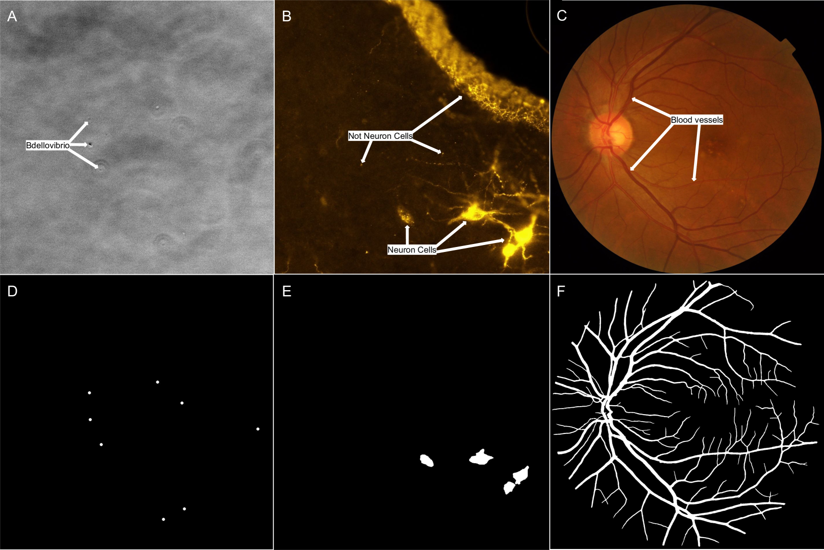
The paper first describes a pipeline for generating biophysically motivated 3D synthetic cell data. This involves modeling the shapes, textures, and spatial arrangements of cells based on physical principles, such as surface tension and diffusion. The synthetic cells are then rendered into 3D volumes that mimic the appearance of real microscopy images.
Next, the researchers evaluated the impact of this synthetic data on the performance of 3D deep learning models for cell segmentation. They trained U-Net models, a common architecture for biomedical image segmentation, using a combination of real and synthetic training data. The models were then tested on held-out real images to measure segmentation accuracy and robustness.
The results showed that incorporating the biophysically motivated synthetic data led to significant improvements in both accuracy and robustness compared to using only real training data. The synthetic data helped the models generalize better and perform more reliably on diverse cell types and imaging conditions.
Critical Analysis
The paper presents a compelling approach for leveraging synthetic data to enhance 3D deep learning for cell segmentation. The biophysical modeling of cell properties is a strength, as it helps ensure the synthetic data is realistic and diverse enough to benefit the training process.
However, the paper does not address potential limitations or caveats of this approach. For example, the synthetic data may not fully capture all the nuances of real microscopy images, such as noise, artifacts, and rare cell types. Additionally, the reliance on synthetic data could make the models more susceptible to distributional shift if the real-world data changes over time.
Further research could explore ways to combine synthetic and real data more effectively, such as through techniques like domain adaptation. Investigating the generalizability of this approach to other 3D biomedical imaging tasks would also be valuable.
Conclusion
This paper presents a promising approach for improving 3D deep learning segmentation of cells through the use of biophysically motivated synthetic data. By generating realistic 3D cell models and incorporating them into the training process, the researchers were able to significantly boost the accuracy and robustness of their cell segmentation models.
This work highlights the potential for synthetic data to complement limited real-world datasets and advance the state-of-the-art in 3D biomedical image analysis. The biophysical modeling techniques developed in this paper could have broader applications in other domains that rely on 3D imaging, such as neuroscience, developmental biology, and medicine.
This summary was produced with help from an AI and may contain inaccuracies - check out the links to read the original source documents!
Related Papers
0
Improving 3D deep learning segmentation with biophysically motivated cell synthesis
Roman Bruch, Mario Vitacolonna, Elina Nurnberg, Simeon Sauer, Rudiger Rudolf, Markus Reischl
Biomedical research increasingly relies on 3D cell culture models and AI-based analysis can potentially facilitate a detailed and accurate feature extraction on a single-cell level. However, this requires for a precise segmentation of 3D cell datasets, which in turn demands high-quality ground truth for training. Manual annotation, the gold standard for ground truth data, is too time-consuming and thus not feasible for the generation of large 3D training datasets. To address this, we present a novel framework for generating 3D training data, which integrates biophysical modeling for realistic cell shape and alignment. Our approach allows the in silico generation of coherent membrane and nuclei signals, that enable the training of segmentation models utilizing both channels for improved performance. Furthermore, we present a new GAN training scheme that generates not only image data but also matching labels. Quantitative evaluation shows superior performance of biophysical motivated synthetic training data, even outperforming manual annotation and pretrained models. This underscores the potential of incorporating biophysical modeling for enhancing synthetic training data quality.
Read more8/30/2024

0
An expert-driven data generation pipeline for histological images
Roberto Basla, Loris Giulivi, Luca Magri, Giacomo Boracchi
Deep Learning (DL) models have been successfully applied to many applications including biomedical cell segmentation and classification in histological images. These models require large amounts of annotated data which might not always be available, especially in the medical field where annotations are scarce and expensive. To overcome this limitation, we propose a novel pipeline for generating synthetic datasets for cell segmentation. Given only a handful of annotated images, our method generates a large dataset of images which can be used to effectively train DL instance segmentation models. Our solution is designed to generate cells of realistic shapes and placement by allowing experts to incorporate domain knowledge during the generation of the dataset.
Read more6/4/2024
0
Perspectives: Comparison of Deep Learning Segmentation Models on Biophysical and Biomedical Data
J Shepard Bryan IV, Meyam Tavakoli, Steve Presse
Deep learning based approaches are now widely used across biophysics to help automate a variety of tasks including image segmentation, feature selection, and deconvolution. However, the presence of multiple competing deep learning architectures, each with its own unique advantages and disadvantages, makes it challenging to select an architecture best suited for a specific application. As such, we present a comprehensive comparison of common models. Here, we focus on the task of segmentation assuming the typically small training dataset sizes available from biophysics experiments and compare the following four commonly used architectures: convolutional neural networks, U-Nets, vision transformers, and vision state space models. In doing so, we establish criteria for determining optimal conditions under which each model excels, thereby offering practical guidelines for researchers and practitioners in the field.
Read more8/16/2024
0
Synthetic Data for Robust Stroke Segmentation
Liam Chalcroft, Ioannis Pappas, Cathy J. Price, John Ashburner
Deep learning-based semantic segmentation in neuroimaging currently requires high-resolution scans and extensive annotated datasets, posing significant barriers to clinical applicability. We present a novel synthetic framework for the task of lesion segmentation, extending the capabilities of the established SynthSeg approach to accommodate large heterogeneous pathologies with lesion-specific augmentation strategies. Our method trains deep learning models, demonstrated here with the UNet architecture, using label maps derived from healthy and stroke datasets, facilitating the segmentation of both healthy tissue and pathological lesions without sequence-specific training data. Evaluated against in-domain and out-of-domain (OOD) datasets, our framework demonstrates robust performance, rivaling current methods within the training domain and significantly outperforming them on OOD data. This contribution holds promise for advancing medical imaging analysis in clinical settings, especially for stroke pathology, by enabling reliable segmentation across varied imaging sequences with reduced dependency on large annotated corpora. Code and weights available at https://github.com/liamchalcroft/SynthStroke.
Read more4/3/2024