Perspectives: Comparison of Deep Learning Segmentation Models on Biophysical and Biomedical Data

0
Sign in to get full access
Overview
- The paper compares the performance of different deep learning models for image segmentation tasks on biophysical and biomedical data.
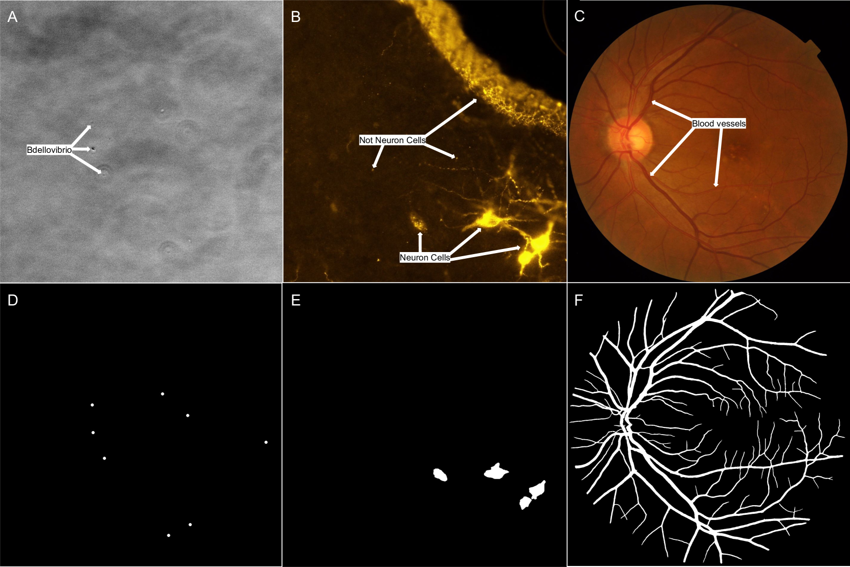
- It evaluates the models on several datasets, including brain MRI, retinal images, and PET-CT scans.
- The goal is to provide insights into the strengths and weaknesses of these models for various medical imaging applications.
Plain English Explanation
The paper looks at how well different deep learning models can segment, or separate, important structures and features in medical images. It tested these models on a variety of datasets, including brain MRI scans, images of the back of the eye, and combined PET-CT scans of the body.
The researchers wanted to understand which deep learning models work best for different medical imaging tasks. This could help doctors and researchers choose the right model for their specific needs, whether that's identifying tumors, blood vessels, or other structures in the body.
The paper provides a side-by-side comparison of the strengths and limitations of these deep learning models. This can guide medical professionals in selecting the most appropriate model for their particular application and help advance the use of AI in healthcare.
Technical Explanation
The paper evaluates the performance of several popular deep learning segmentation models, including U-Net, Mask R-CNN, and DeepLabV3+, on a range of biophysical and biomedical datasets. These datasets include:
- Brain MRI dataset for segmenting brain tissues
- Retinal blood vessel dataset for segmenting vasculature in the eye
- PET-CT tumor dataset for segmenting tumors in combined PET-CT scans
The models are evaluated on metrics such as Dice score, Jaccard index, and pixel-wise accuracy to assess their segmentation performance. The results show that different models excel in different medical imaging tasks, with no single "best" model across all datasets.
Critical Analysis
The paper provides a comprehensive and objective comparison of deep learning segmentation models on biophysical and biomedical data. However, it acknowledges several limitations and areas for future work:
- The datasets used may not be representative of all real-world medical imaging scenarios, and model performance may vary on other datasets.
- The paper does not explore the computational efficiency or inference speed of the models, which are also important factors in practical medical applications.
- The analysis could be expanded to include more segmentation models, including newer architectural variants and ensemble methods.
Additionally, the paper does not delve into the interpretability and explainability of these deep learning models, which is an important consideration for their adoption in clinical settings.
Conclusion
This paper offers a valuable comparative analysis of deep learning segmentation models on biophysical and biomedical data. The findings can guide medical professionals in selecting the most appropriate model for their specific imaging tasks, while also highlighting areas for further research and development in this rapidly evolving field.
The insights provided can contribute to the broader adoption of AI-powered tools in healthcare, ultimately improving diagnostic accuracy, treatment planning, and patient outcomes.
This summary was produced with help from an AI and may contain inaccuracies - check out the links to read the original source documents!
Related Papers
0
Perspectives: Comparison of Deep Learning Segmentation Models on Biophysical and Biomedical Data
J Shepard Bryan IV, Meyam Tavakoli, Steve Presse
Deep learning based approaches are now widely used across biophysics to help automate a variety of tasks including image segmentation, feature selection, and deconvolution. However, the presence of multiple competing deep learning architectures, each with its own unique advantages and disadvantages, makes it challenging to select an architecture best suited for a specific application. As such, we present a comprehensive comparison of common models. Here, we focus on the task of segmentation assuming the typically small training dataset sizes available from biophysics experiments and compare the following four commonly used architectures: convolutional neural networks, U-Nets, vision transformers, and vision state space models. In doing so, we establish criteria for determining optimal conditions under which each model excels, thereby offering practical guidelines for researchers and practitioners in the field.
Read more8/16/2024

0
Improving 3D deep learning segmentation with biophysically motivated cell synthesis
Roman Bruch, Mario Vitacolonna, Elina Nurnberg, Simeon Sauer, Rudiger Rudolf, Markus Reischl
Biomedical research increasingly relies on 3D cell culture models and AI-based analysis can potentially facilitate a detailed and accurate feature extraction on a single-cell level. However, this requires for a precise segmentation of 3D cell datasets, which in turn demands high-quality ground truth for training. Manual annotation, the gold standard for ground truth data, is too time-consuming and thus not feasible for the generation of large 3D training datasets. To address this, we present a novel framework for generating 3D training data, which integrates biophysical modeling for realistic cell shape and alignment. Our approach allows the in silico generation of coherent membrane and nuclei signals, that enable the training of segmentation models utilizing both channels for improved performance. Furthermore, we present a new GAN training scheme that generates not only image data but also matching labels. Quantitative evaluation shows superior performance of biophysical motivated synthetic training data, even outperforming manual annotation and pretrained models. This underscores the potential of incorporating biophysical modeling for enhancing synthetic training data quality.
Read more8/30/2024

0
Biomedical Image Segmentation: A Systematic Literature Review of Deep Learning Based Object Detection Methods
Fazli Wahid, Yingliang Ma, Dawar Khan, Muhammad Aamir, Syed U. K. Bukhari
Biomedical image segmentation plays a vital role in diagnosis of diseases across various organs. Deep learning-based object detection methods are commonly used for such segmentation. There exists an extensive research in this topic. However, there is no standard review on this topic. Existing surveys often lack a standardized approach or focus on broader segmentation techniques. In this paper, we conducted a systematic literature review (SLR), collected and analysed 148 articles that explore deep learning object detection methods for biomedical image segmentation. We critically analyzed these methods, identified the key challenges, and discussed the future directions. From the selected articles we extracted the results including the deep learning models, targeted imaging modalities, targeted diseases, and the metrics for the analysis of the methods. The results have been presented in tabular and/or charted forms. The results are presented in three major categories including two stage detection models, one stage detection models and point-based detection models. Each article is individually analyzed along with its pros and cons. Finally, we discuss open challenges, potential benefits, and future research directions. This SLR aims to provide the research community with a quick yet deeper understanding of these segmentation models, ultimately facilitating the development of more powerful solutions for biomedical image analysis.
Read more8/30/2024

0
TBConvL-Net: A Hybrid Deep Learning Architecture for Robust Medical Image Segmentation
Shahzaib Iqbal, Tariq M. Khan, Syed S. Naqvi, Asim Naveed, Erik Meijering
Deep learning has shown great potential for automated medical image segmentation to improve the precision and speed of disease diagnostics. However, the task presents significant difficulties due to variations in the scale, shape, texture, and contrast of the pathologies. Traditional convolutional neural network (CNN) models have certain limitations when it comes to effectively modelling multiscale context information and facilitating information interaction between skip connections across levels. To overcome these limitations, a novel deep learning architecture is introduced for medical image segmentation, taking advantage of CNNs and vision transformers. Our proposed model, named TBConvL-Net, involves a hybrid network that combines the local features of a CNN encoder-decoder architecture with long-range and temporal dependencies using biconvolutional long-short-term memory (LSTM) networks and vision transformers (ViT). This enables the model to capture contextual channel relationships in the data and account for the uncertainty of segmentation over time. Additionally, we introduce a novel composite loss function that considers both the segmentation robustness and the boundary agreement of the predicted output with the gold standard. Our proposed model shows consistent improvement over the state of the art on ten publicly available datasets of seven different medical imaging modalities.
Read more9/6/2024