Improving Deep Learning-based Automatic Cranial Defect Reconstruction by Heavy Data Augmentation: From Image Registration to Latent Diffusion Models

0
Sign in to get full access
Overview
- This paper presents a deep learning-based approach for automatically reconstructing cranial defects using heavy data augmentation techniques, including image registration and latent diffusion models.
- The researchers aim to improve the performance of deep learning models for this task, which has important applications in craniofacial surgery and reconstruction.
- The paper covers various techniques, from traditional image registration to more advanced latent diffusion models, to generate diverse and realistic training data for the deep learning models.
Plain English Explanation
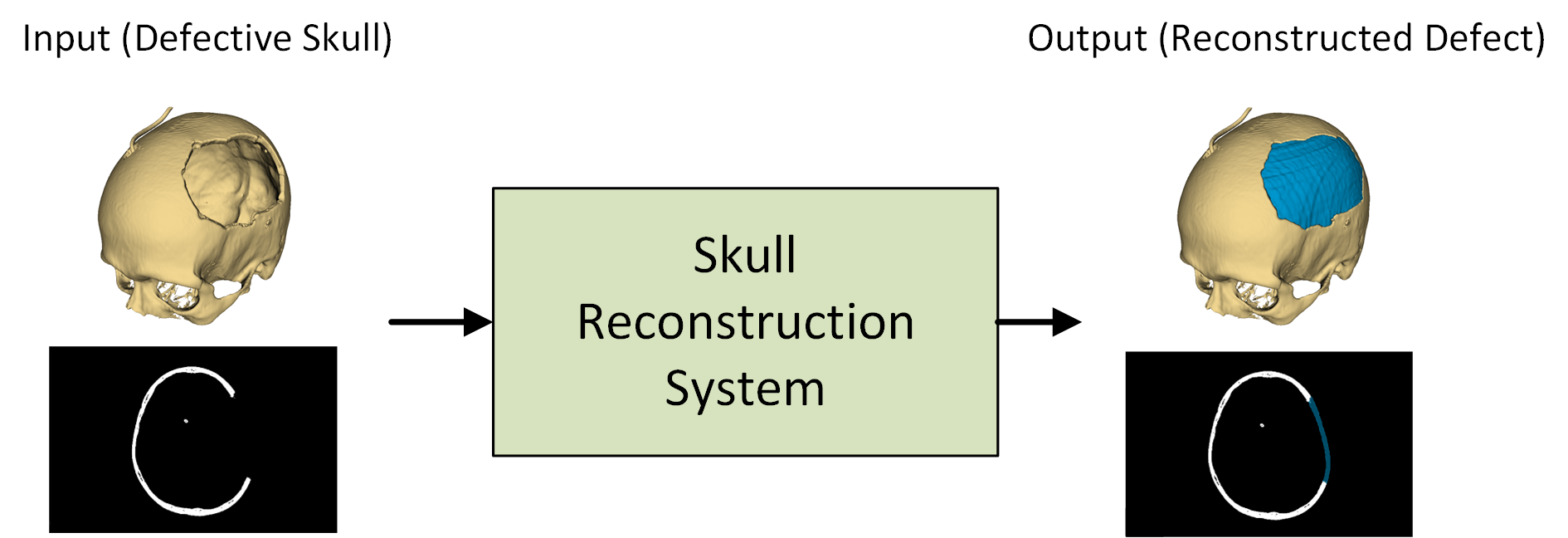
The human skull can sometimes have defects or abnormalities, such as holes or misshapen areas. These cranial defects can occur due to injury, disease, or congenital conditions. Repairing these defects is an important task in craniofacial surgery, and deep learning models have shown promise in automating this process.
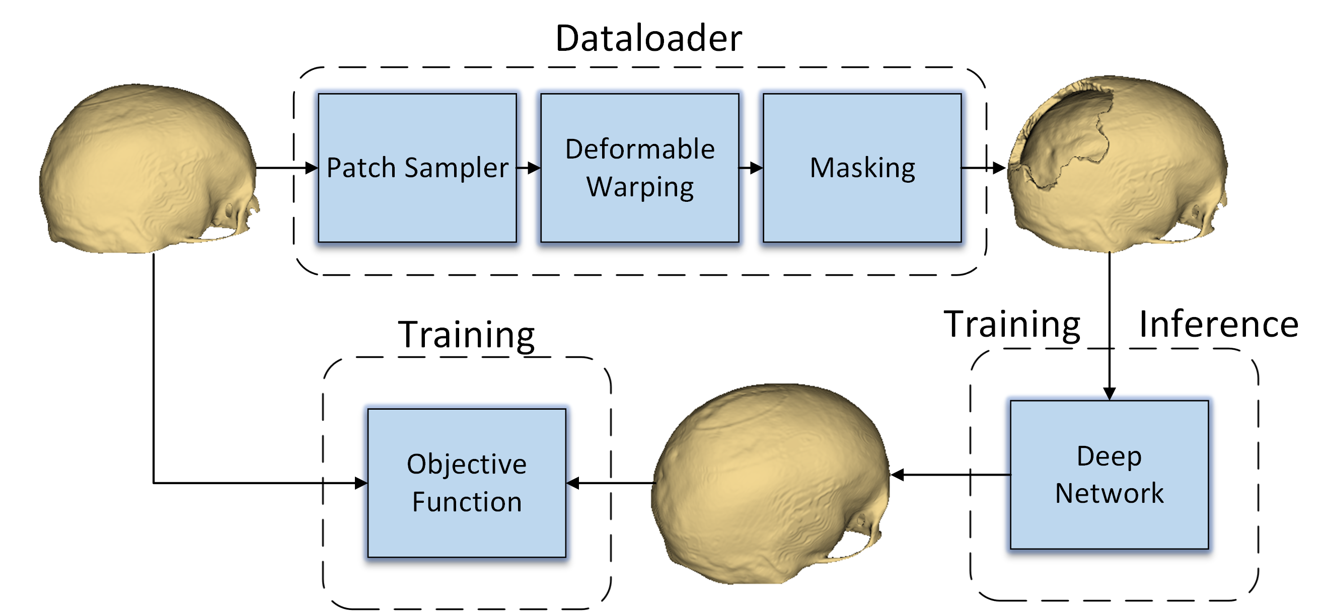
However, training deep learning models for cranial defect reconstruction can be challenging due to the limited availability of high-quality training data. This paper explores techniques to address this problem by generating synthetic training data through heavy data augmentation.
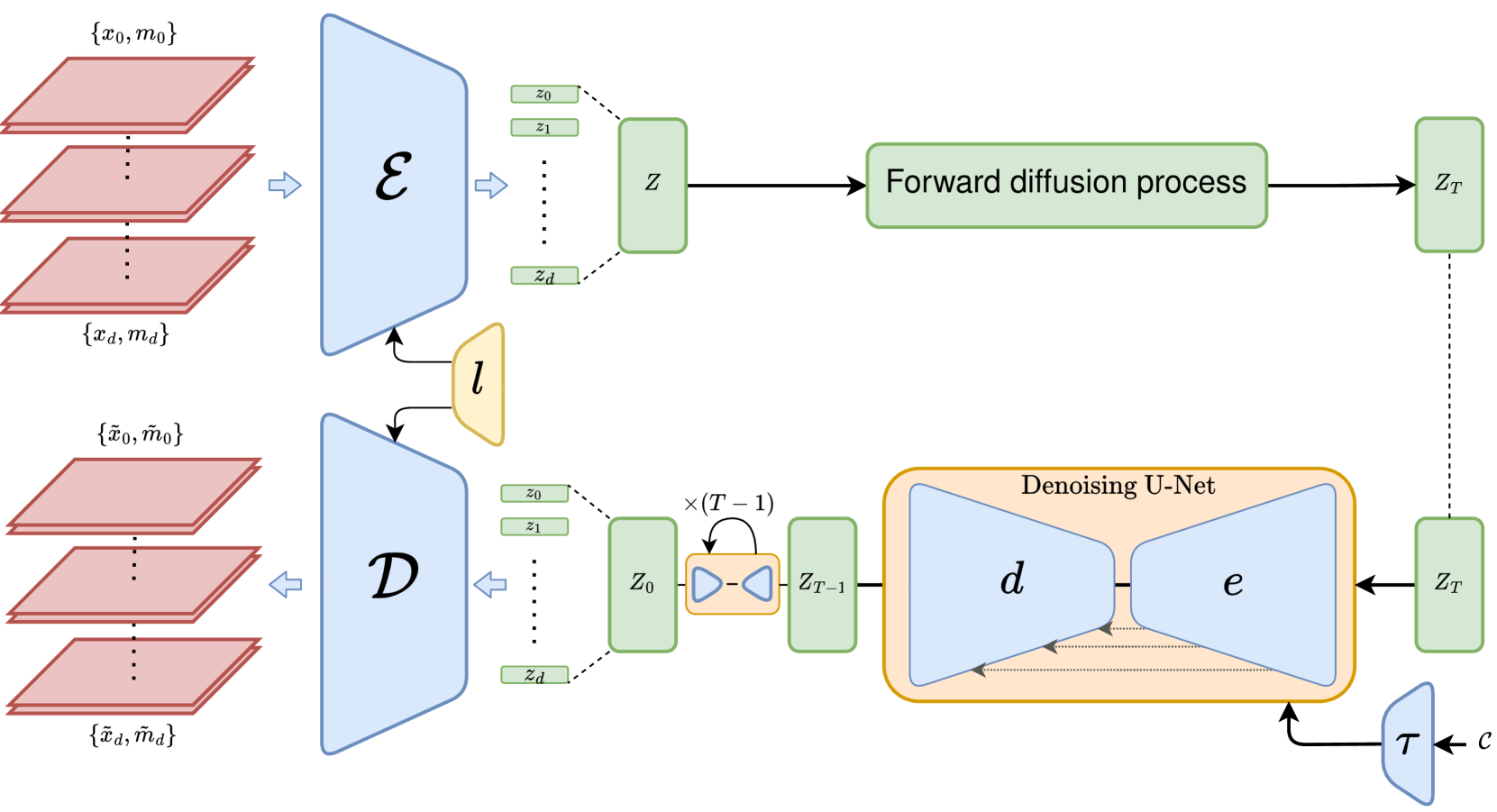
The researchers start with traditional image registration methods, which can align and transform existing cranial images to create new variations. They then move on to more advanced latent diffusion models, which can generate completely novel, realistic-looking cranial images with defects. Latent diffusion models are a type of generative AI that learns to create new images by understanding the underlying patterns in a dataset.
By combining these data augmentation techniques, the researchers were able to significantly improve the performance of deep learning models for automatically reconstructing cranial defects. This could lead to more accurate and efficient tools for craniofacial surgeons, ultimately benefiting patients with these types of medical conditions.
Technical Explanation
The paper presents a deep learning-based approach for automatic cranial defect reconstruction, focusing on improving model performance through heavy data augmentation techniques.
The researchers first explore traditional image registration methods, which can be used to align and transform existing cranial images to create new variations for training. This includes techniques like affine and deformable registration, which can introduce realistic geometric and intensity variations to the training data.
Building on this, the paper then introduces the use of latent diffusion models for cranial defect reconstruction. These generative AI models can create completely novel, high-quality cranial images with realistic defects by learning the underlying patterns in the training data. The researchers leverage diffusion-based image generation techniques to train the latent diffusion model and generate diverse training samples.
The deep learning model for cranial defect reconstruction is trained on the augmented dataset, which includes both registered and diffusion-generated images. The researchers evaluate the model's performance on various metrics, such as reconstruction accuracy and visual realism, and compare it to previous approaches for this task.
Critical Analysis
The paper presents a comprehensive approach to improving deep learning-based cranial defect reconstruction, leveraging both traditional and state-of-the-art data augmentation techniques. The use of latent diffusion models is particularly noteworthy, as it allows for the generation of highly realistic and diverse training samples that can significantly boost model performance.
However, the paper does acknowledge some limitations of the proposed approach. For example, the latent diffusion model may struggle to capture the full complexity of cranial anatomy, and the generated samples may not perfectly match the characteristics of real-world defects. Additionally, the paper does not provide a detailed analysis of the computational cost and training time required for the data augmentation and model training processes.
Further research could explore ways to address these limitations, such as incorporating more domain-specific knowledge into the latent diffusion model or developing efficient training strategies. Additionally, 3D-based cranial defect reconstruction and the use of diffusion-based deepfake generation techniques could be interesting avenues for future investigation.
Overall, this paper presents a promising approach to enhancing deep learning-based cranial defect reconstruction, with the potential to improve surgical planning and outcomes for patients with these types of craniofacial conditions.
Conclusion
This paper introduces a deep learning-based approach for automatic cranial defect reconstruction that leverages heavy data augmentation techniques, including traditional image registration and more advanced latent diffusion models. By generating diverse and realistic training data, the researchers were able to significantly improve the performance of the deep learning models for this task.
The use of latent diffusion models is a particularly noteworthy contribution, as it allows for the creation of novel, high-quality cranial images with realistic defects. This approach could have far-reaching implications for craniofacial surgery and reconstruction, potentially leading to more accurate and efficient tools for surgeons to plan and execute complex procedures.
While the paper acknowledges some limitations, the overall findings demonstrate the power of data augmentation and generative AI in addressing the challenges of limited training data in medical imaging applications. As the field of deep learning continues to evolve, techniques like those presented in this paper will likely play an increasingly important role in advancing the state of the art in various healthcare domains.
This summary was produced with help from an AI and may contain inaccuracies - check out the links to read the original source documents!
Related Papers
0
Improving Deep Learning-based Automatic Cranial Defect Reconstruction by Heavy Data Augmentation: From Image Registration to Latent Diffusion Models
Marek Wodzinski, Kamil Kwarciak, Mateusz Daniol, Daria Hemmerling
Modeling and manufacturing of personalized cranial implants are important research areas that may decrease the waiting time for patients suffering from cranial damage. The modeling of personalized implants may be partially automated by the use of deep learning-based methods. However, this task suffers from difficulties with generalizability into data from previously unseen distributions that make it difficult to use the research outcomes in real clinical settings. Due to difficulties with acquiring ground-truth annotations, different techniques to improve the heterogeneity of datasets used for training the deep networks have to be considered and introduced. In this work, we present a large-scale study of several augmentation techniques, varying from classical geometric transformations, image registration, variational autoencoders, and generative adversarial networks, to the most recent advances in latent diffusion models. We show that the use of heavy data augmentation significantly increases both the quantitative and qualitative outcomes, resulting in an average Dice Score above 0.94 for the SkullBreak and above 0.96 for the SkullFix datasets. Moreover, we show that the synthetically augmented network successfully reconstructs real clinical defects. The work is a considerable contribution to the field of artificial intelligence in the automatic modeling of personalized cranial implants.
Read more6/11/2024
0
Automatic Cranial Defect Reconstruction with Self-Supervised Deep Deformable Masked Autoencoders
Marek Wodzinski, Daria Hemmerling, Mateusz Daniol
Thousands of people suffer from cranial injuries every year. They require personalized implants that need to be designed and manufactured before the reconstruction surgery. The manual design is expensive and time-consuming leading to searching for algorithms whose goal is to automatize the process. The problem can be formulated as volumetric shape completion and solved by deep neural networks dedicated to supervised image segmentation. However, such an approach requires annotating the ground-truth defects which is costly and time-consuming. Usually, the process is replaced with synthetic defect generation. However, even the synthetic ground-truth generation is time-consuming and limits the data heterogeneity, thus the deep models' generalizability. In our work, we propose an alternative and simple approach to use a self-supervised masked autoencoder to solve the problem. This approach by design increases the heterogeneity of the training set and can be seen as a form of data augmentation. We compare the proposed method with several state-of-the-art deep neural networks and show both the quantitative and qualitative improvement on the SkullBreak and SkullFix datasets. The proposed method can be used to efficiently reconstruct the cranial defects in real time.
Read more6/4/2024
🚀
0
High-Resolution Cranial Defect Reconstruction by Iterative, Low-Resolution, Point Cloud Completion Transformers
Marek Wodzinski, Mateusz Daniol, Daria Hemmerling, Miroslaw Socha
Each year thousands of people suffer from various types of cranial injuries and require personalized implants whose manual design is expensive and time-consuming. Therefore, an automatic, dedicated system to increase the availability of personalized cranial reconstruction is highly desirable. The problem of the automatic cranial defect reconstruction can be formulated as the shape completion task and solved using dedicated deep networks. Currently, the most common approach is to use the volumetric representation and apply deep networks dedicated to image segmentation. However, this approach has several limitations and does not scale well into high-resolution volumes, nor takes into account the data sparsity. In our work, we reformulate the problem into a point cloud completion task. We propose an iterative, transformer-based method to reconstruct the cranial defect at any resolution while also being fast and resource-efficient during training and inference. We compare the proposed methods to the state-of-the-art volumetric approaches and show superior performance in terms of GPU memory consumption while maintaining high-quality of the reconstructed defects.
Read more5/22/2024
0
3D MRI Synthesis with Slice-Based Latent Diffusion Models: Improving Tumor Segmentation Tasks in Data-Scarce Regimes
Aghiles Kebaili, J'er^ome Lapuyade-Lahorgue, Pierre Vera, Su Ruan
Despite the increasing use of deep learning in medical image segmentation, the limited availability of annotated training data remains a major challenge due to the time-consuming data acquisition and privacy regulations. In the context of segmentation tasks, providing both medical images and their corresponding target masks is essential. However, conventional data augmentation approaches mainly focus on image synthesis. In this study, we propose a novel slice-based latent diffusion architecture designed to address the complexities of volumetric data generation in a slice-by-slice fashion. This approach extends the joint distribution modeling of medical images and their associated masks, allowing a simultaneous generation of both under data-scarce regimes. Our approach mitigates the computational complexity and memory expensiveness typically associated with diffusion models. Furthermore, our architecture can be conditioned by tumor characteristics, including size, shape, and relative position, thereby providing a diverse range of tumor variations. Experiments on a segmentation task using the BRATS2022 confirm the effectiveness of the synthesized volumes and masks for data augmentation.
Read more6/11/2024