Improving the Scan-rescan Precision of AI-based CMR Biomarker Estimation

0

Sign in to get full access
Overview
- The paper explores ways to improve the precision of AI-based cardiac biomarker estimation from cardiac magnetic resonance (CMR) imaging.
- Key focus is on reducing the variability in biomarker measurements when the same patient is scanned multiple times (scan-rescan precision).
- Researchers developed an AI-based model and tested it on a dataset of CMR scans from multiple visits.
Plain English Explanation
The paper looks at how to make the measurements of certain health indicators, called biomarkers, more consistent when using AI to analyze cardiac MRI scans. Biomarkers are important for assessing heart health, but the measurements can vary when the same person gets scanned multiple times. The researchers wanted to find ways to reduce this variability, or "scan-rescan" inconsistency, in the AI-based analysis.
They created an AI model and tested it on a dataset of cardiac MRI scans from patients who had multiple scans over time. The goal was to see if they could develop an AI approach that would provide more reliable and consistent biomarker measurements, even when scanning the same person multiple times.
Technical Explanation
The researchers developed an AI-based model for estimating cardiac biomarkers from CMR images. To address the scan-rescan variability issue, they explored several technical approaches:
-
Data Augmentation: They used data augmentation techniques like image flipping, scaling, and rotation to artificially expand the training dataset and improve the model's ability to generalize.
-
Domain Adaptation: The model was trained to adapt to differences in scanner types, imaging protocols, and patient populations across the training and test datasets.
-
Ensemble Modeling: Multiple AI models were trained and their outputs were combined to produce more robust and stable biomarker estimates.
The model was evaluated on a dataset of CMR scans from 100 patients who had 2-3 scans each. The scan-rescan precision of the AI-based biomarker estimates was compared to manual expert analysis.
Critical Analysis
The paper provides a thorough technical approach to improving the reliability of AI-based cardiac biomarker estimation. However, some limitations and areas for further research are worth noting:
-
The dataset, while sizeable, may not fully capture the diversity of patient populations and scanner types seen in real-world clinical practice. Further validation on larger and more heterogeneous datasets is needed.
-
The paper does not explore the impact of model interpretability or explainability, which could be important for building clinical trust in the AI system.
-
The scan-rescan precision improvements, while significant, still leave room for further optimization. Exploring additional techniques like self-supervised learning or federated learning may yield even better results.
Conclusion
This research represents an important step towards making AI-based cardiac biomarker estimation more reliable and consistent, which is crucial for its widespread clinical adoption. The techniques developed, such as data augmentation and ensemble modeling, provide a framework for improving scan-rescan precision that could be applicable to other medical imaging domains as well. Continued research in this area has the potential to enhance the accuracy and trustworthiness of AI-powered diagnostic tools for cardiac care.
This summary was produced with help from an AI and may contain inaccuracies - check out the links to read the original source documents!
Related Papers
0
Improving the Scan-rescan Precision of AI-based CMR Biomarker Estimation
Dewmini Hasara Wickremasinghe, Yiyang Xu, Esther Puyol-Ant'on, Paul Aljabar, Reza Razavi, Andrew P. King
Quantification of cardiac biomarkers from cine cardiovascular magnetic resonance (CMR) data using deep learning (DL) methods offers many advantages, such as increased accuracy and faster analysis. However, only a few studies have focused on the scan-rescan precision of the biomarker estimates, which is important for reproducibility and longitudinal analysis. Here, we propose a cardiac biomarker estimation pipeline that not only focuses on achieving high segmentation accuracy but also on improving the scan-rescan precision of the computed biomarkers, namely left and right ventricular ejection fraction, and left ventricular myocardial mass. We evaluate two approaches to improve the apical-basal resolution of the segmentations used for estimating the biomarkers: one based on image interpolation and one based on segmentation interpolation. Using a database comprising scan-rescan cine CMR data acquired from 92 subjects, we compare the performance of these two methods against ground truth (GT) segmentations and DL segmentations obtained before interpolation (baseline). The results demonstrate that both the image-based and segmentation-based interpolation methods were able to narrow Bland-Altman scan-rescan confidence intervals for all biomarkers compared to the GT and baseline performances. Our findings highlight the importance of focusing not only on segmentation accuracy but also on the consistency of biomarkers across repeated scans, which is crucial for longitudinal analysis of cardiac function.
Read more8/22/2024
🤿
0
Inline AI: Open-source Deep Learning Inference for Cardiac MR
Hui Xue, Rhodri H Davies, James Howard, Hunain Shiwani, Azaan Rehman, Iain Pierce, Henry Procter, Marianna Fontana, James C Moon, Eylem Levelt, Peter Kellman
Cardiac Magnetic Resonance (CMR) is established as a non-invasive imaging technique for evaluation of heart function, anatomy, and myocardial tissue characterization. Quantitative biomarkers are central for diagnosis and management of heart disease. Deep learning (DL) is playing an ever more important role in extracting these quantitative measures from CMR images. While many researchers have reported promising results in training and evaluating models, model deployment into the imaging workflow is less explored. A new imaging AI framework, the InlineAI, was developed and open-sourced. The main innovation is to enable the model inference inline as a part of imaging computation, instead of as an offline post-processing step and to allow users to plug in their models. We demonstrate the system capability on three applications: long-axis CMR cine landmark detection, short-axis CMR cine analysis of function and anatomy, and quantitative perfusion mapping. The InlineAI allowed models to be deployed into imaging workflow in a streaming manner directly on the scanner. The model was loaded and inference on incoming images were performed while the data acquisition was ongoing, and results were sent back to scanner. Several biomarkers were extracted from model outputs in the demonstrated applications and reported as curves and tabular values. All processes are full automated. the model inference was completed within 6-45s after the end of imaging data acquisition.
Read more4/4/2024
🎯
0
Temporal-spatial Adaptation of Promptable SAM Enhance Accuracy and Generalizability of cine CMR Segmentation
Zhennong Chen, Sekeun Kim, Hui Ren, Quanzheng Li, Xiang Li
Accurate myocardium segmentation across all phases in one cardiac cycle in cine cardiac magnetic resonance (CMR) scans is crucial for comprehensively cardiac function analysis. Despite advancements in deep learning (DL) for automatic cine CMR segmentation, generalizability on unseen data remains a significant challenge. Recently, the segment-anything-model (SAM) has been invented as a segmentation foundation model, known for its accurate segmentation and more importantly, zero-shot generalization. SAM was trained on two-dimensional (2D) natural images; to adapt it for comprehensive cine CMR segmentation, we propose cineCMR-SAM which incorporates both temporal and spatial information through a modified model architecture. Compared to other state-of-the-art (SOTA) methods, our model achieved superior data-specific model segmentation accuracy on the STACOM2011 when fine-tuned on this dataset and demonstrated superior zero-shot generalization on two other large public datasets (ACDC and M&Ms) unseen during fine-tuning. Additionally, we introduced a text prompt feature in cineCMR-SAM to specify the view type of input slices (short-axis or long-axis), enhancing performance across all view types.
Read more7/17/2024
0
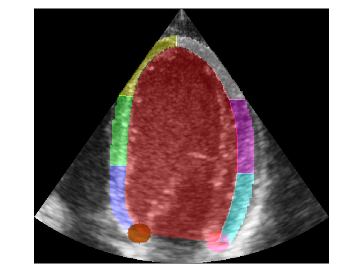
Regional quality estimation for echocardiography using deep learning
Gilles Van De Vyver, Svein-Erik M{aa}s{o}y, H{aa}vard Dalen, Bj{o}rnar Leangen Grenne, Espen Holte, Sindre Hellum Olaisen, John Nyberg, Andreas {O}stvik, Lasse L{o}vstakken, Erik Smistad
Automatic estimation of cardiac ultrasound image quality can be beneficial for guiding operators and ensuring the accuracy of clinical measurements. Previous work often fails to distinguish the view correctness of the echocardiogram from the image quality. Additionally, previous studies only provide a global image quality value, which limits their practical utility. In this work, we developed and compared three methods to estimate image quality: 1) classic pixel-based metrics like the generalized contrast-to-noise ratio (gCNR) on myocardial segments as region of interest and left ventricle lumen as background, obtained using a U-Net segmentation 2) local image coherence derived from a U-Net model that predicts coherence from B-Mode images 3) a deep convolutional network that predicts the quality of each region directly in an end-to-end fashion. We evaluate each method against manual regional image quality annotations by three experienced cardiologists. The results indicate poor performance of the gCNR metric, with Spearman correlation to the annotations of rho = 0.24. The end-to-end learning model obtains the best result, rho = 0.69, comparable to the inter-observer correlation, rho = 0.63. Finally, the coherence-based method, with rho = 0.58, outperformed the classical metrics and is more generic than the end-to-end approach.
Read more8/28/2024