Regional quality estimation for echocardiography using deep learning

0
Sign in to get full access
Overview
- This paper presents a deep learning-based approach for estimating the quality of different regions in echocardiography images.
- Echocardiography is a widely used medical imaging technique that captures images of the heart, but the quality of these images can vary.
- The proposed method aims to automatically assess the quality of different regions within echocardiography images to help clinicians identify areas that may require further examination.
Plain English Explanation
Echocardiography is a type of medical imaging that uses sound waves to create pictures of the heart. These images can be very helpful for doctors to diagnose and monitor heart conditions. However, the quality of these images can sometimes be uneven, with some areas being clearer than others.
The researchers in this study developed a new deep learning algorithm that can automatically evaluate the quality of different regions within echocardiography images. Deep learning is a type of artificial intelligence that can identify patterns in data, like images, and learn to make predictions or decisions.
The idea is that this algorithm could help doctors quickly identify parts of the echocardiography image that may need a closer look or might be harder to interpret. This could make the diagnostic process more efficient and accurate, ultimately leading to better care for patients with heart problems.
Technical Explanation
The researchers trained a deep learning model to assess the quality of different regions within echocardiography images. They used a convolutional neural network (CNN) architecture, which is well-suited for analyzing image data.
The model was trained on a large dataset of echocardiography images, where each image was manually annotated by experts to indicate the quality of different regions. The model learned to recognize the visual patterns associated with high-quality and low-quality regions.
During inference, the trained model can then be applied to new echocardiography images to automatically generate a "quality map" that highlights the regions with high and low quality. This information could be used by clinicians to focus their attention on the most reliable parts of the image when making diagnoses or treatment decisions.
Critical Analysis
The researchers acknowledge several limitations of their approach. First, the dataset used for training was relatively small, which may limit the model's generalization to a wider range of echocardiography images. Expanding the dataset with more diverse examples could improve the model's performance.
Additionally, the study only evaluated the model's ability to assess quality at the regional level, but did not assess its impact on downstream clinical decision-making. Further research is needed to understand how this technology could be integrated into clinical workflows and its effect on patient outcomes.
It's also worth noting that the quality of echocardiography images can be influenced by a variety of factors, such as the patient's anatomy, the skill of the technician performing the scan, and the specific equipment used. The deep learning model may not be able to account for all of these nuances, and its reliability may vary depending on the clinical setting.
Conclusion
This research demonstrates the potential of deep learning to automatically assess the quality of echocardiography images at the regional level. By highlighting areas of high and low quality, this technology could help clinicians focus their attention on the most reliable parts of the image, potentially leading to more accurate diagnoses and better patient care.
However, further research is needed to expand the dataset, evaluate the model's impact on clinical decision-making, and understand its limitations in real-world settings. As with any new technology, it will be important to carefully assess the benefits and risks before widespread adoption in clinical practice.
This summary was produced with help from an AI and may contain inaccuracies - check out the links to read the original source documents!
Related Papers
0
Regional quality estimation for echocardiography using deep learning
Gilles Van De Vyver, Svein-Erik M{aa}s{o}y, H{aa}vard Dalen, Bj{o}rnar Leangen Grenne, Espen Holte, Sindre Hellum Olaisen, John Nyberg, Andreas {O}stvik, Lasse L{o}vstakken, Erik Smistad
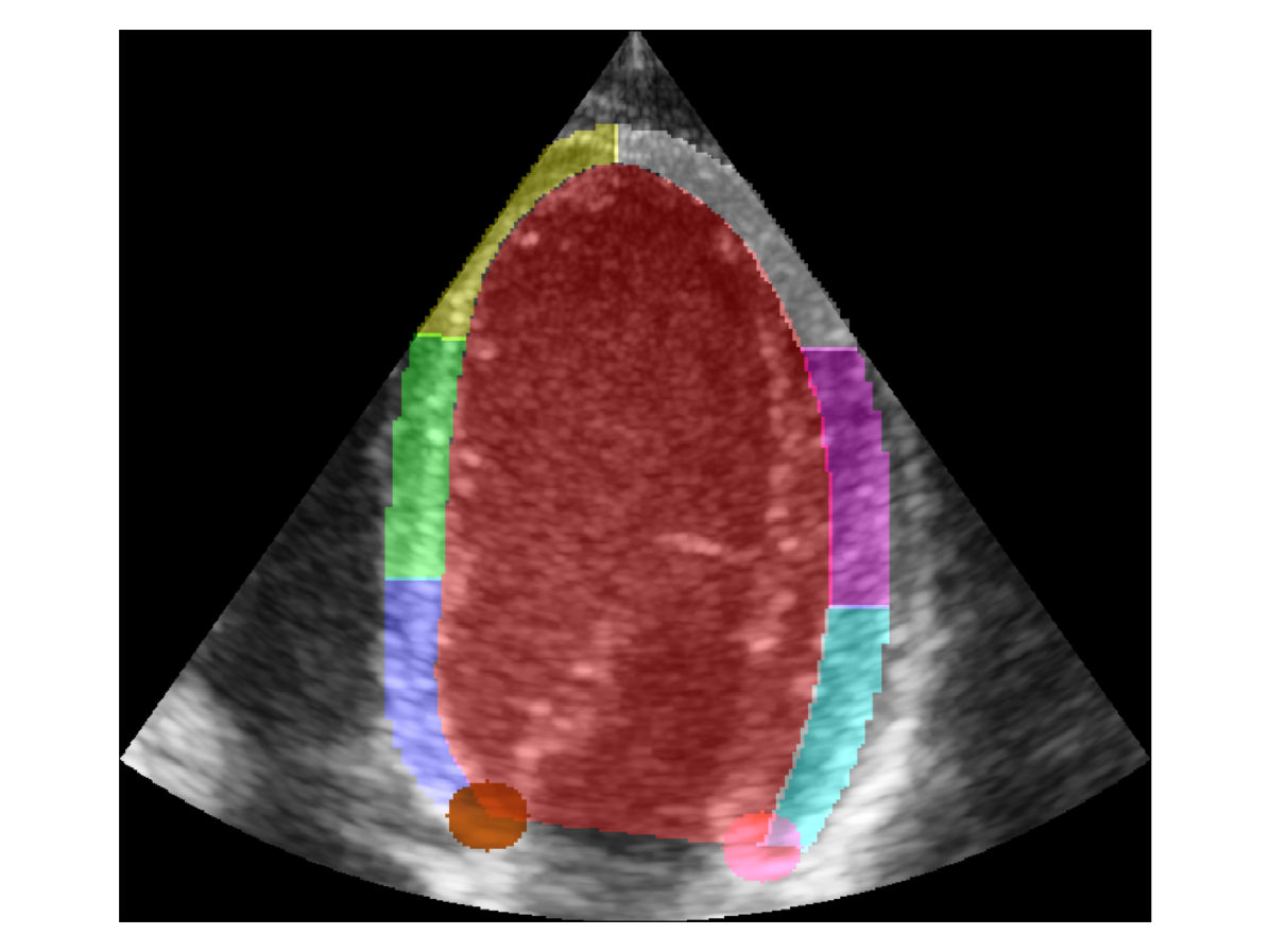
Automatic estimation of cardiac ultrasound image quality can be beneficial for guiding operators and ensuring the accuracy of clinical measurements. Previous work often fails to distinguish the view correctness of the echocardiogram from the image quality. Additionally, previous studies only provide a global image quality value, which limits their practical utility. In this work, we developed and compared three methods to estimate image quality: 1) classic pixel-based metrics like the generalized contrast-to-noise ratio (gCNR) on myocardial segments as region of interest and left ventricle lumen as background, obtained using a U-Net segmentation 2) local image coherence derived from a U-Net model that predicts coherence from B-Mode images 3) a deep convolutional network that predicts the quality of each region directly in an end-to-end fashion. We evaluate each method against manual regional image quality annotations by three experienced cardiologists. The results indicate poor performance of the gCNR metric, with Spearman correlation to the annotations of rho = 0.24. The end-to-end learning model obtains the best result, rho = 0.69, comparable to the inter-observer correlation, rho = 0.63. Finally, the coherence-based method, with rho = 0.58, outperformed the classical metrics and is more generic than the end-to-end approach.
Read more8/28/2024

0
Ultrafast Cardiac Imaging Using Deep Learning For Speckle-Tracking Echocardiography
Jingfeng Lu, Fabien Millioz, Franc{c}ois Varray, Jonathan Por'ee, Jean Provost, Olivier Bernard, Damien Garcia, Denis Friboulet
High-quality ultrafast ultrasound imaging is based on coherent compounding from multiple transmissions of plane waves (PW) or diverging waves (DW). However, compounding results in reduced frame rate, as well as destructive interferences from high-velocity tissue motion if motion compensation (MoCo) is not considered. While many studies have recently shown the interest of deep learning for the reconstruction of high-quality static images from PW or DW, its ability to achieve such performance while maintaining the capability of tracking cardiac motion has yet to be assessed. In this paper, we addressed such issue by deploying a complex-weighted convolutional neural network (CNN) for image reconstruction and a state-of-the-art speckle tracking method. The evaluation of this approach was first performed by designing an adapted simulation framework, which provides specific reference data, i.e. high quality, motion artifact-free cardiac images. The obtained results showed that, while using only three DWs as input, the CNN-based approach yielded an image quality and a motion accuracy equivalent to those obtained by compounding 31 DWs free of motion artifacts. The performance was then further evaluated on non-simulated, experimental in vitro data, using a spinning disk phantom. This experiment demonstrated that our approach yielded high-quality image reconstruction and motion estimation, under a large range of velocities and outperforms a state-of-the-art MoCo-based approach at high velocities. Our method was finally assessed on in vivo datasets and showed consistent improvement in image quality and motion estimation compared to standard compounding. This demonstrates the feasibility and effectiveness of deep learning reconstruction for ultrafast speckle-tracking echocardiography.
Read more7/30/2024

0
Improving the Scan-rescan Precision of AI-based CMR Biomarker Estimation
Dewmini Hasara Wickremasinghe, Yiyang Xu, Esther Puyol-Ant'on, Paul Aljabar, Reza Razavi, Andrew P. King
Quantification of cardiac biomarkers from cine cardiovascular magnetic resonance (CMR) data using deep learning (DL) methods offers many advantages, such as increased accuracy and faster analysis. However, only a few studies have focused on the scan-rescan precision of the biomarker estimates, which is important for reproducibility and longitudinal analysis. Here, we propose a cardiac biomarker estimation pipeline that not only focuses on achieving high segmentation accuracy but also on improving the scan-rescan precision of the computed biomarkers, namely left and right ventricular ejection fraction, and left ventricular myocardial mass. We evaluate two approaches to improve the apical-basal resolution of the segmentations used for estimating the biomarkers: one based on image interpolation and one based on segmentation interpolation. Using a database comprising scan-rescan cine CMR data acquired from 92 subjects, we compare the performance of these two methods against ground truth (GT) segmentations and DL segmentations obtained before interpolation (baseline). The results demonstrate that both the image-based and segmentation-based interpolation methods were able to narrow Bland-Altman scan-rescan confidence intervals for all biomarkers compared to the GT and baseline performances. Our findings highlight the importance of focusing not only on segmentation accuracy but also on the consistency of biomarkers across repeated scans, which is crucial for longitudinal analysis of cardiac function.
Read more8/22/2024
0
Boosting Cardiac Color Doppler Frame Rates with Deep Learning
Julia Puig, Denis Friboulet, Hang Jung Ling, Franc{c}ois Varray, Jonathan Por'ee, Jean Provost, Damien Garcia, Fabien Millioz
Color Doppler echocardiography enables visualization of blood flow within the heart. However, the limited frame rate impedes the quantitative assessment of blood velocity throughout the cardiac cycle, thereby compromising a comprehensive analysis of ventricular filling. Concurrently, deep learning is demonstrating promising outcomes in post-processing of echocardiographic data for various applications. This work explores the use of deep learning models for intracardiac Doppler velocity estimation from a reduced number of filtered I/Q signals. We used a supervised learning approach by simulating patient-based cardiac color Doppler acquisitions and proposed data augmentation strategies to enlarge the training dataset. We implemented architectures based on convolutional neural networks. In particular, we focused on comparing the U-Net model and the recent ConvNeXt models, alongside assessing real-valued versus complex-valued representations. We found that both models outperformed the state-of-the-art autocorrelator method, effectively mitigating aliasing and noise. We did not observe significant differences between the use of real and complex data. Finally, we validated the models on in vitro and in vivo experiments. All models produced quantitatively comparable results to the baseline and were more robust to noise. ConvNeXt emerged as the sole model to achieve high-quality results on in vivo aliased samples. These results demonstrate the interest of supervised deep learning methods for Doppler velocity estimation from a reduced number of acquisitions.
Read more8/22/2024