Integrated Brain Connectivity Analysis with fMRI, DTI, and sMRI Powered by Interpretable Graph Neural Networks

0
🧠
Sign in to get full access
Overview
- The provided paper discusses a novel approach for generating brain connectivity graphs from diffusion magnetic resonance imaging (dMRI) data using adversarial deep learning.
- The researchers developed a generative adversarial network (GAN) model to transform dMRI data into realistic brain connectivity graphs, which can be used for a variety of applications in neuroscience and clinical research.
- The paper presents the model architecture, training process, and evaluation of the generated graphs compared to ground-truth data.
Plain English Explanation
The human brain is a complex network of interconnected neurons that communicate with each other. Researchers often study these brain connections, or "connectivity," using a technique called diffusion magnetic resonance imaging (dMRI). This method can capture the movement of water molecules in the brain, which provides information about the underlying structure of the brain's wiring.
However, interpreting and analyzing dMRI data can be challenging. The researchers in this paper developed a new way to address this problem. They created a machine learning model based on a generative adversarial network (GAN). This type of model learns to generate realistic-looking brain connectivity graphs from the dMRI data.
The key idea is that the model learns to produce connectivity graphs that are indistinguishable from real ones, by competing with another part of the model that tries to identify the fake graphs. Through this "adversarial" training process, the generator part of the model becomes better and better at generating highly realistic brain connectivity graphs.
These generated graphs can then be used for a variety of applications, such as studying brain development, diagnosing neurological disorders, or even simulating brain function. The advantage of this approach is that it can produce a large number of realistic brain connectivity graphs from a relatively small amount of dMRI data, which can be valuable for researchers and clinicians.
Technical Explanation
The researchers developed a generative adversarial network (GAN) model to transform diffusion magnetic resonance imaging (dMRI) data into realistic brain connectivity graphs. The model consists of two main components:
- Generator: This part of the model learns to generate brain connectivity graphs that resemble the ground-truth graphs derived from the dMRI data.
- Discriminator: The discriminator attempts to distinguish the generated graphs from the real ones, providing feedback to the generator to improve its performance.
The training process involves iteratively updating the generator and discriminator, with the goal of the generator producing graphs that the discriminator cannot reliably identify as fake. This adversarial training allows the generator to learn the underlying structure and characteristics of the real brain connectivity graphs.
To evaluate the model, the researchers compared the generated graphs to the ground-truth graphs using several metrics, such as graph-level statistics (e.g., edge density, clustering coefficient) and node-level measures (e.g., degree distribution, centrality). The results showed that the generated graphs closely matched the real ones, demonstrating the effectiveness of the GAN-based approach.
Critical Analysis
The researchers acknowledge several limitations and areas for further investigation:
-
Interpretability: While the generated graphs closely resemble the real ones, the internal workings of the GAN model may not be fully interpretable. Establishing a better understanding of how the model learns to generate the graphs could be valuable for gaining insights into brain connectivity.
-
Generalization: The study was conducted on a specific dataset, and the researchers note that further evaluation is needed to assess the model's performance on more diverse dMRI data, including data from different brain imaging modalities or clinical populations.
-
Evaluation Metrics: The researchers used a set of established graph-level and node-level metrics to evaluate the generated graphs, but there may be other relevant measures that could provide additional insights into the quality and plausibility of the generated graphs.
-
Clinical Applications: While the researchers discuss potential applications in neuroscience and clinical research, the paper does not provide a detailed analysis of how the generated graphs could be used in real-world settings, such as for disease diagnosis or treatment planning. Further research is needed to explore the practical implications of this approach.
Overall, the paper presents a promising approach for generating realistic brain connectivity graphs from dMRI data using adversarial deep learning. However, the interpretation, generalization, and clinical applications of the method warrant further investigation to fully understand its potential impact in the field of neuroscience and healthcare.
Conclusion
This paper introduces a novel GAN-based approach for transforming diffusion magnetic resonance imaging (dMRI) data into realistic brain connectivity graphs. The generated graphs closely match the statistical properties of the ground-truth data, demonstrating the effectiveness of the adversarial training process.
The ability to generate large, diverse sets of brain connectivity graphs from limited dMRI data has the potential to significantly impact various areas of neuroscience and clinical research, such as studying brain development, diagnosing neurological disorders, and simulating brain function. While the work presents some limitations and areas for further exploration, it represents an important step forward in leveraging the power of deep learning to gain deeper insights into the complex structure and dynamics of the human brain.
This summary was produced with help from an AI and may contain inaccuracies - check out the links to read the original source documents!
Related Papers
🧠
0
Integrated Brain Connectivity Analysis with fMRI, DTI, and sMRI Powered by Interpretable Graph Neural Networks
Gang Qu, Ziyu Zhou, Vince D. Calhoun, Aiying Zhang, Yu-Ping Wang
Multimodal neuroimaging modeling has becomes a widely used approach but confronts considerable challenges due to heterogeneity, which encompasses variability in data types, scales, and formats across modalities. This variability necessitates the deployment of advanced computational methods to integrate and interpret these diverse datasets within a cohesive analytical framework. In our research, we amalgamate functional magnetic resonance imaging, diffusion tensor imaging, and structural MRI into a cohesive framework. This integration capitalizes on the unique strengths of each modality and their inherent interconnections, aiming for a comprehensive understanding of the brain's connectivity and anatomical characteristics. Utilizing the Glasser atlas for parcellation, we integrate imaging derived features from various modalities: functional connectivity from fMRI, structural connectivity from DTI, and anatomical features from sMRI within consistent regions. Our approach incorporates a masking strategy to differentially weight neural connections, thereby facilitating a holistic amalgamation of multimodal imaging data. This technique enhances interpretability at connectivity level, transcending traditional analyses centered on singular regional attributes. The model is applied to the Human Connectome Project's Development study to elucidate the associations between multimodal imaging and cognitive functions throughout youth. The analysis demonstrates improved predictive accuracy and uncovers crucial anatomical features and essential neural connections, deepening our understanding of brain structure and function.
Read more8/27/2024
🛸
0
Brain Imaging-to-Graph Generation using Adversarial Hierarchical Diffusion Models for MCI Causality Analysis
Qiankun Zuo, Hao Tian, Chi-Man Pun, Hongfei Wang, Yudong Zhang, Jin Hong
Effective connectivity can describe the causal patterns among brain regions. These patterns have the potential to reveal the pathological mechanism and promote early diagnosis and effective drug development for cognitive disease. However, the current methods utilize software toolkits to extract empirical features from brain imaging to estimate effective connectivity. These methods heavily rely on manual parameter settings and may result in large errors during effective connectivity estimation. In this paper, a novel brain imaging-to-graph generation (BIGG) framework is proposed to map functional magnetic resonance imaging (fMRI) into effective connectivity for mild cognitive impairment (MCI) analysis. To be specific, the proposed BIGG framework is based on the diffusion denoising probabilistic models (DDPM), where each denoising step is modeled as a generative adversarial network (GAN) to progressively translate the noise and conditional fMRI to effective connectivity. The hierarchical transformers in the generator are designed to estimate the noise at multiple scales. Each scale concentrates on both spatial and temporal information between brain regions, enabling good quality in noise removal and better inference of causal relations. Meanwhile, the transformer-based discriminator constrains the generator to further capture global and local patterns for improving high-quality and diversity generation. By introducing the diffusive factor, the denoising inference with a large sampling step size is more efficient and can maintain high-quality results for effective connectivity generation. Evaluations of the ADNI dataset demonstrate the feasibility and efficacy of the proposed model. The proposed model not only achieves superior prediction performance compared with other competing methods but also predicts MCI-related causal connections that are consistent with clinical studies.
Read more6/4/2024
↗️
0
Diffusion MRI with Machine Learning
Davood Karimi
Diffusion-weighted magnetic resonance imaging (dMRI) offers unique capabilities including noninvasive probing of brain's tissue microstructure and structural connectivity. It is widely used for clinical assessment of brain pathologies and for neuroscience research. Analyzing the dMRI data to extract useful information for medical and scientific purposes can be challenging. The dMRI measurements often suffer from strong noise and artifacts, there is usually high inter-session and inter-scanner variability in the data, and considerable inter-subject heterogeneity in brain structure. Moreover, the relationship between measurements and the phenomena of interest can be highly complex. Recent years have witnessed increasing use of machine learning methods for dMRI analysis. This manuscript aims to assess these efforts, with a focus on methods that have addressed data preprocessing and harmonization, microstructure mapping, tractography, and white matter tract analysis. We study the main findings, strengths, and weaknesses of the existing methods and suggest topics for future research. We find that machine learning may be exceptionally suited to tackle some of the difficult tasks in dMRI analysis. However, for this to happen, several shortcomings of existing methods and critical unresolved issues need to be addressed. These include deficient evaluation practices, lack of rich training datasets and validation benchmarks, as well as model generalizability, reliability, and explainability concerns.
Read more7/29/2024
0
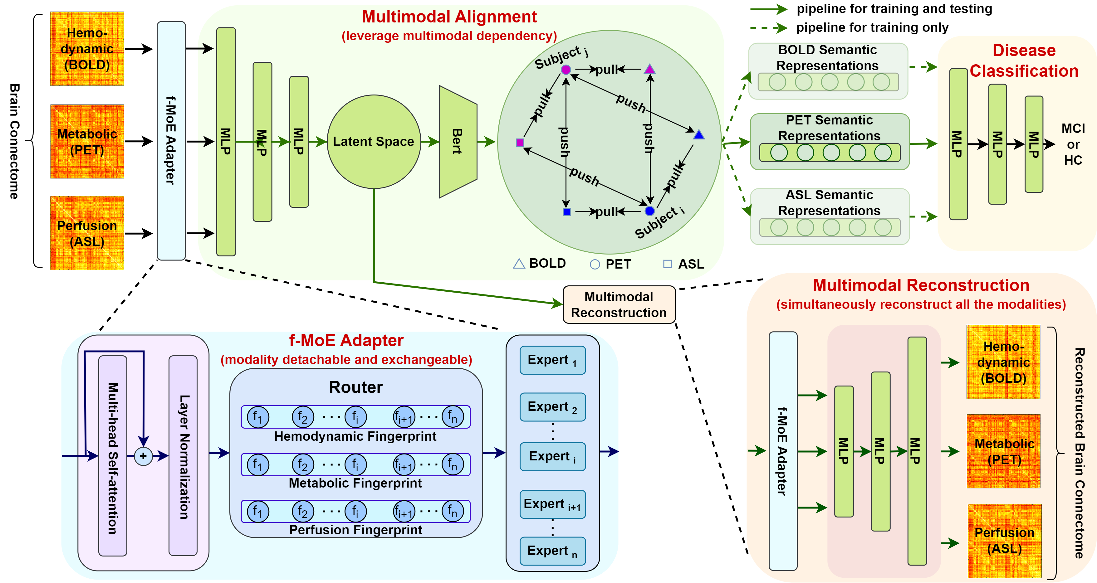
Revolutionizing Disease Diagnosis with simultaneous functional PET/MR and Deeply Integrated Brain Metabolic, Hemodynamic, and Perfusion Networks
Luoyu Wang, Yitian Tao, Qing Yang, Yan Liang, Siwei Liu, Hongcheng Shi, Dinggang Shen, Han Zhang
Simultaneous functional PET/MR (sf-PET/MR) presents a cutting-edge multimodal neuroimaging technique. It provides an unprecedented opportunity for concurrently monitoring and integrating multifaceted brain networks built by spatiotemporally covaried metabolic activity, neural activity, and cerebral blood flow (perfusion). Albeit high scientific/clinical values, short in hardware accessibility of PET/MR hinders its applications, let alone modern AI-based PET/MR fusion models. Our objective is to develop a clinically feasible AI-based disease diagnosis model trained on comprehensive sf-PET/MR data with the power of, during inferencing, allowing single modality input (e.g., PET only) as well as enforcing multimodal-based accuracy. To this end, we propose MX-ARM, a multimodal MiXture-of-experts Alignment and Reconstruction Model. It is modality detachable and exchangeable, allocating different multi-layer perceptrons dynamically (mixture of experts) through learnable weights to learn respective representations from different modalities. Such design will not sacrifice model performance in uni-modal situation. To fully exploit the inherent complex and nonlinear relation among modalities while producing fine-grained representations for uni-modal inference, we subsequently add a modal alignment module to line up a dominant modality (e.g., PET) with representations of auxiliary modalities (MR). We further adopt multimodal reconstruction to promote the quality of learned features. Experiments on precious multimodal sf-PET/MR data for Mild Cognitive Impairment diagnosis showcase the efficacy of our model toward clinically feasible precision medicine.
Read more4/1/2024