Revolutionizing Disease Diagnosis with simultaneous functional PET/MR and Deeply Integrated Brain Metabolic, Hemodynamic, and Perfusion Networks
2403.20058
0
0
Abstract
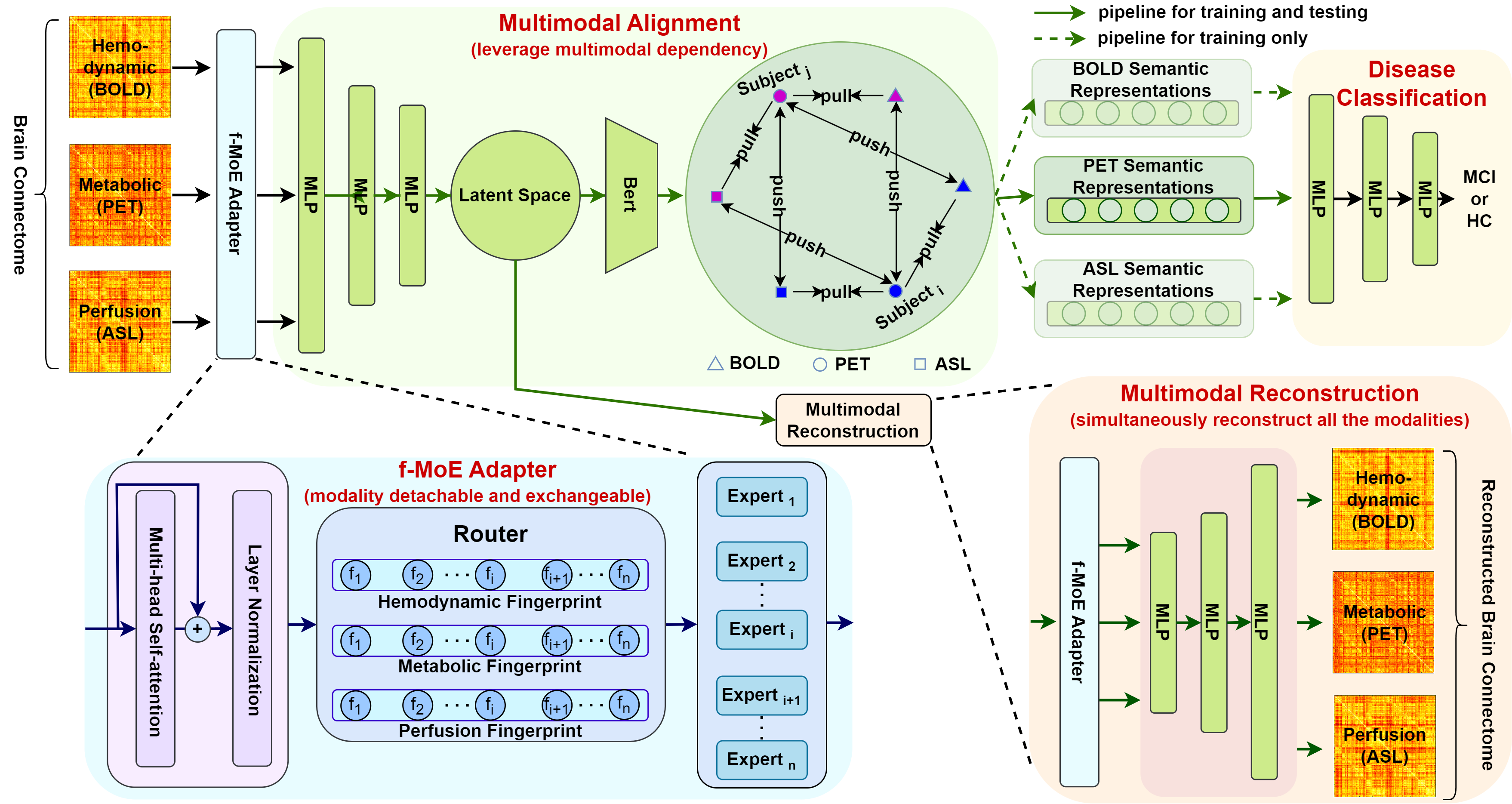
Simultaneous functional PET/MR (sf-PET/MR) presents a cutting-edge multimodal neuroimaging technique. It provides an unprecedented opportunity for concurrently monitoring and integrating multifaceted brain networks built by spatiotemporally covaried metabolic activity, neural activity, and cerebral blood flow (perfusion). Albeit high scientific/clinical values, short in hardware accessibility of PET/MR hinders its applications, let alone modern AI-based PET/MR fusion models. Our objective is to develop a clinically feasible AI-based disease diagnosis model trained on comprehensive sf-PET/MR data with the power of, during inferencing, allowing single modality input (e.g., PET only) as well as enforcing multimodal-based accuracy. To this end, we propose MX-ARM, a multimodal MiXture-of-experts Alignment and Reconstruction Model. It is modality detachable and exchangeable, allocating different multi-layer perceptrons dynamically (mixture of experts) through learnable weights to learn respective representations from different modalities. Such design will not sacrifice model performance in uni-modal situation. To fully exploit the inherent complex and nonlinear relation among modalities while producing fine-grained representations for uni-modal inference, we subsequently add a modal alignment module to line up a dominant modality (e.g., PET) with representations of auxiliary modalities (MR). We further adopt multimodal reconstruction to promote the quality of learned features. Experiments on precious multimodal sf-PET/MR data for Mild Cognitive Impairment diagnosis showcase the efficacy of our model toward clinically feasible precision medicine.
Create account to get full access
Overview
- This paper presents a novel approach to disease diagnosis using simultaneous functional Positron Emission Tomography (PET) and Magnetic Resonance Imaging (MRI) scans, as well as a deep analysis of integrated brain metabolic, hemodynamic, and perfusion networks.
- The key focus is on revolutionizing the early diagnosis of Alzheimer's disease (AD) through this multimodal neuroimaging technique.
- The researchers aim to provide a more comprehensive understanding of brain function and connectivity, which could lead to earlier detection and intervention for various neurological disorders.
Plain English Explanation
The paper describes a new medical imaging technique that combines two powerful tools - PET and MRI scans. PET scans measure the activity and metabolism of different brain regions, while MRI scans provide detailed structural information about the brain. By using these two technologies together, the researchers can get a more complete picture of how the brain is functioning.
This is particularly useful for diagnosing conditions like Alzheimer's disease, which affect the brain in complex ways. Traditional methods of detecting Alzheimer's may not catch the disease until it has progressed quite far. But by looking at both the metabolic changes and the structural changes in the brain, the researchers believe they can identify Alzheimer's much earlier, before significant damage has occurred.
The key innovation is the "deep integration" of the different types of brain data - the metabolic, hemodynamic (blood flow), and perfusion (tissue absorption) information. By analyzing these interconnected brain networks in-depth, the researchers hope to uncover new insights that could revolutionize how we detect and understand neurodegenerative diseases like Alzheimer's.
Technical Explanation
The core of the research is the simultaneous acquisition and analysis of functional PET and MRI data. PET scans measure the brain's metabolic activity by tracking the uptake of radioactive tracers, while MRI provides high-resolution structural and functional imaging of the brain.
By aligning and integrating these multimodal datasets, the researchers can gain a more comprehensive view of brain physiology. They use advanced machine learning techniques to model the complex interactions between metabolic, hemodynamic, and perfusion-based brain networks. This allows them to identify patterns that may be early indicators of Alzheimer's disease and other neurological disorders.
The experimental design involved scanning participants with both PET and MRI, then using computational methods to spatially and temporally register the images. This enabled the researchers to extract features like regional metabolic rates, cerebral blood flow, and tissue perfusion. They then applied deep learning models to analyze the relationships between these integrated brain measures.
The key technical insights include the discovery of disruptions to the functional connectivity of metabolic, hemodynamic, and perfusion networks in Alzheimer's patients, as compared to healthy controls. These network-level changes appear to precede the structural brain atrophy typically associated with AD, suggesting the potential for earlier diagnosis.
Critical Analysis
The paper presents a compelling technical approach to multimodal neuroimaging analysis, with a clear focus on improving early detection of Alzheimer's disease. The simultaneous PET/MRI data acquisition and deep integration of complementary brain measures is a notable methodological advancement.
However, the research is still in an early stage, with relatively small sample sizes and a need for further validation on independent patient cohorts. The authors acknowledge these limitations and indicate the need for larger-scale longitudinal studies to fully assess the clinical utility of this approach.
Additionally, while the deep learning techniques are powerful, there remains a need for more interpretable and explainable models that can provide insights into the underlying neurobiology. The "black box" nature of some deep neural networks may hinder the clinical adoption of these methods.
It would also be valuable to explore how this integrated brain imaging approach could be applied to the diagnosis and monitoring of other neurological and psychiatric disorders, beyond just Alzheimer's disease. Demonstrating the broader applicability of the techniques would strengthen the overall impact and significance of the research.
Conclusion
This paper presents a novel multimodal neuroimaging technique that combines simultaneous PET and MRI data to provide a more holistic view of brain function. The deep integration of metabolic, hemodynamic, and perfusion-based brain networks shows promise for revolutionizing the early detection of Alzheimer's disease and potentially other neurological disorders.
While the research is still in an early phase, the technical advances and insights gained could have far-reaching implications for the field of disease diagnosis and monitoring. Continued development and validation of these methods may lead to earlier interventions, improved patient outcomes, and a better understanding of the complex neurobiological mechanisms underlying devastating neurodegenerative conditions.
This summary was produced with help from an AI and may contain inaccuracies - check out the links to read the original source documents!
Related Papers
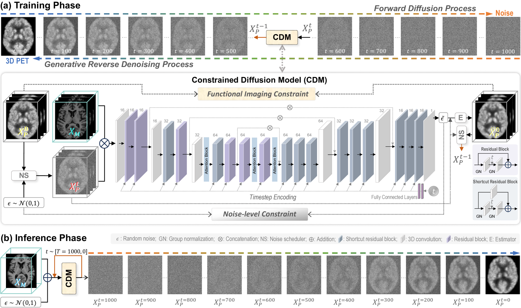
Functional Imaging Constrained Diffusion for Brain PET Synthesis from Structural MRI
Minhui Yu, Mengqi Wu, Ling Yue, Andrea Bozoki, Mingxia Liu
0
0
Magnetic resonance imaging (MRI) and positron emission tomography (PET) are increasingly used in multimodal analysis of neurodegenerative disorders. While MRI is broadly utilized in clinical settings, PET is less accessible. Many studies have attempted to use deep generative models to synthesize PET from MRI scans. However, they often suffer from unstable training and inadequately preserve brain functional information conveyed by PET. To this end, we propose a functional imaging constrained diffusion (FICD) framework for 3D brain PET image synthesis with paired structural MRI as input condition, through a new constrained diffusion model (CDM). The FICD introduces noise to PET and then progressively removes it with CDM, ensuring high output fidelity throughout a stable training phase. The CDM learns to predict denoised PET with a functional imaging constraint introduced to ensure voxel-wise alignment between each denoised PET and its ground truth. Quantitative and qualitative analyses conducted on 293 subjects with paired T1-weighted MRI and 18F-fluorodeoxyglucose (FDG)-PET scans suggest that FICD achieves superior performance in generating FDG-PET data compared to state-of-the-art methods. We further validate the effectiveness of the proposed FICD on data from a total of 1,262 subjects through three downstream tasks, with experimental results suggesting its utility and generalizability.
5/10/2024
🤯
Three-Dimensional Amyloid-Beta PET Synthesis from Structural MRI with Conditional Generative Adversarial Networks
Fernando Vega, Abdoljalil Addeh, M. Ethan MacDonald
0
0
Motivation: Alzheimer's Disease hallmarks include amyloid-beta deposits and brain atrophy, detectable via PET and MRI scans, respectively. PET is expensive, invasive and exposes patients to ionizing radiation. MRI is cheaper, non-invasive, and free from ionizing radiation but limited to measuring brain atrophy. Goal: To develop an 3D image translation model that synthesizes amyloid-beta PET images from T1-weighted MRI, exploiting the known relationship between amyloid-beta and brain atrophy. Approach: The model was trained on 616 PET/MRI pairs and validated with 264 pairs. Results: The model synthesized amyloid-beta PET images from T1-weighted MRI with high-degree of similarity showing high SSIM and PSNR metrics (SSIM>0.95&PSNR=28). Impact: Our model proves the feasibility of synthesizing amyloid-beta PET images from structural MRI ones, significantly enhancing accessibility for large-cohort studies and early dementia detection, while also reducing cost, invasiveness, and radiation exposure.
5/6/2024

PASTA: Pathology-Aware MRI to PET Cross-Modal Translation with Diffusion Models
Yitong Li, Igor Yakushev, Dennis M. Hedderich, Christian Wachinger
0
0
Positron emission tomography (PET) is a well-established functional imaging technique for diagnosing brain disorders. However, PET's high costs and radiation exposure limit its widespread use. In contrast, magnetic resonance imaging (MRI) does not have these limitations. Although it also captures neurodegenerative changes, MRI is a less sensitive diagnostic tool than PET. To close this gap, we aim to generate synthetic PET from MRI. Herewith, we introduce PASTA, a novel pathology-aware image translation framework based on conditional diffusion models. Compared to the state-of-the-art methods, PASTA excels in preserving both structural and pathological details in the target modality, which is achieved through its highly interactive dual-arm architecture and multi-modal condition integration. A cycle exchange consistency and volumetric generation strategy elevate PASTA's capability to produce high-quality 3D PET scans. Our qualitative and quantitative results confirm that the synthesized PET scans from PASTA not only reach the best quantitative scores but also preserve the pathology correctly. For Alzheimer's classification, the performance of synthesized scans improves over MRI by 4%, almost reaching the performance of actual PET. Code is available at https://github.com/ai-med/PASTA.
5/28/2024

Multimodal MRI-based Detection of Amyloid Status in Alzheimer's Disease Continuum
Giorgio Dolci (Department of Computer Science, University of Verona, Verona, Italy, Department of Engineering for Innovation Medicine, University of Verona, Verona, Italy, Tri-Institutional Center for Translational Research in Neuroimaging and Data Science), Charles A. Ellis (Tri-Institutional Center for Translational Research in Neuroimaging and Data Science), Federica Cruciani (Department of Engineering for Innovation Medicine, University of Verona, Verona, Italy), Lorenza Brusini (Department of Engineering for Innovation Medicine, University of Verona, Verona, Italy), Anees Abrol (Tri-Institutional Center for Translational Research in Neuroimaging and Data Science), Ilaria Boscolo Galazzo (Department of Engineering for Innovation Medicine, University of Verona, Verona, Italy), Gloria Menegaz (Department of Engineering for Innovation Medicine, University of Verona, Verona, Italy), Vince D. Calhoun (Tri-Institutional Center for Translational Research in Neuroimaging and Data Science)
0
0
Amyloid-$beta$ (A$beta$) plaques in conjunction with hyperphosphorylated tau proteins in the form of neurofibrillary tangles are the two neuropathological hallmarks of Alzheimer's disease (AD). In particular, the accumulation of A$beta$ plaques, as evinced by the A/T/N (amyloid/tau/neurodegeneration) framework, marks the initial stage. Thus, the identification of individuals with A$beta$ positivity could enable early diagnosis and potentially lead to more effective interventions. Deep learning methods relying mainly on amyloid PET images have been employed to this end. However, PET imaging has some disadvantages, including the need of radiotracers and expensive acquisitions. Hence, in this work, we propose a novel multimodal approach that integrates information from structural, functional, and diffusion MRI data to discriminate A$beta$ status in the AD continuum. Our method achieved an accuracy of $0.762pm0.04$. Furthermore, a textit{post-hoc} explainability analysis (guided backpropagation) was performed to retrieve the brain regions that most influenced the model predictions. This analysis identified some key regions that were common across modalities, some of which were well-established AD-discriminative biomarkers and related to A$beta$ deposition, such as the hippocampus, thalamus, precuneus, and cingulate gyrus. Hence, our study demonstrates the potential viability of MRI-based characterization of A$beta$ status, paving the way for further research in this domain.
6/21/2024