LeDNet: Localization-enabled Deep Neural Network for Multi-Label Radiography Image Classification

0
Sign in to get full access
Overview
- This paper presents LeDNet, a deep neural network for multi-label classification of radiography images.
- LeDNet can not only classify the diseases present in an image, but also localize the regions of the image that are relevant to the identified diseases.
- The authors evaluate LeDNet on a dataset of chest X-ray images and show that it outperforms existing state-of-the-art methods in both classification and localization tasks.
Plain English Explanation
The paper introduces LeDNet, which is a deep learning model designed to analyze medical images like chest X-rays. Deep learning is a powerful technique that can automatically learn to recognize patterns in data, and it has shown great promise in computer-aided diagnosis of thoracic diseases from medical images.
What makes LeDNet special is that it can not only classify the diseases present in an image, but also localize the specific regions of the image that are relevant to each identified disease. This is important because it can help doctors understand which parts of the image are contributing to the diagnosis, which can aid in their decision-making process.
The authors evaluate LeDNet on a dataset of chest X-ray images and show that it outperforms existing state-of-the-art methods in both the classification and localization tasks. This suggests that LeDNet could be a valuable tool for diagnosing thoracic diseases from chest X-rays.
Technical Explanation
The authors formulate the problem as a multi-label classification task, where the goal is to identify all the diseases present in a given radiography image. To solve this problem, they propose the LeDNet architecture, which consists of a feature extraction backbone and a classification and localization head.
The feature extraction backbone is based on a convolutional neural network (CNN) that learns to extract relevant visual features from the input image. The classification and localization head then uses these features to predict the diseases present in the image and localize the regions associated with each disease.
The authors train and evaluate LeDNet on a dataset of chest X-ray images, comparing its performance to several state-of-the-art methods. They show that LeDNet achieves superior classification and localization accuracy, demonstrating the effectiveness of their approach.
Critical Analysis
The paper provides a comprehensive evaluation of LeDNet and demonstrates its strong performance on the task of multi-label radiography image classification with localization. However, there are a few potential limitations and areas for further research that could be explored:
-
Dataset Diversity: The authors only evaluate LeDNet on a single dataset of chest X-ray images. It would be valuable to assess the model's performance on a more diverse set of radiography images, such as those from different anatomical regions or modalities, to better understand its generalizability.
-
Interpretability: While LeDNet can localize the relevant regions in the image, the authors do not provide a detailed analysis of the interpretability of these localizations. Further research could explore ways to improve the interpretability of the model's decision-making process.
-
Real-World Deployment: The paper focuses on the technical aspects of LeDNet, but does not address the practical challenges of deploying such a system in a real-world clinical setting. Factors like integration with existing workflows, data privacy, and regulatory considerations would need to be addressed for successful translation to clinical practice.
-
Comparison to Human Performance: The authors compare LeDNet's performance to other machine learning models, but it would be valuable to also assess its performance relative to human experts in radiography interpretation to better understand the model's potential clinical utility.
Conclusion
This paper presents a promising deep learning model, LeDNet, for multi-label classification and localization of radiography images. The authors demonstrate that LeDNet outperforms existing state-of-the-art methods, suggesting its potential as a valuable tool for computer-aided diagnosis of thoracic diseases. While the technical details of the model are well-described, the paper also highlights areas for further research and consideration of practical deployment challenges. Overall, this work represents an important step forward in the development of advanced AI-based tools for medical image analysis.
This summary was produced with help from an AI and may contain inaccuracies - check out the links to read the original source documents!
Related Papers
0
LeDNet: Localization-enabled Deep Neural Network for Multi-Label Radiography Image Classification
Lalit Pant, Shubham Arora
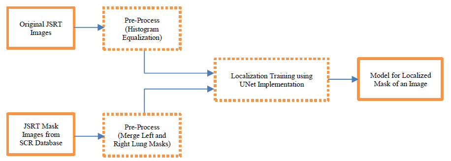
Multi-label radiography image classification has long been a topic of interest in neural networks research. In this paper, we intend to classify such images using convolution neural networks with novel localization techniques. We will use the chest x-ray images to detect thoracic diseases for this purpose. For accurate diagnosis, it is crucial to train the network with good quality images. But many chest X-ray images have irrelevant external objects like distractions created by faulty scans, electronic devices scanned next to lung region, scans inadvertently capturing bodily air etc. To address these, we propose a combination of localization and deep learning algorithms called LeDNet to predict thoracic diseases with higher accuracy. We identify and extract the lung region masks from chest x-ray images through localization. These masks are superimposed on the original X-ray images to create the mask overlay images. DenseNet-121 classification models are then used for feature selection to retrieve features of the entire chest X-ray images and the localized mask overlay images. These features are then used to predict disease classification. Our experiments involve comparing classification results obtained with original CheXpert images and mask overlay images. The comparison is demonstrated through accuracy and loss curve analyses.
Read more7/8/2024

0
InfLocNet: Enhanced Lung Infection Localization and Disease Detection from Chest X-Ray Images Using Lightweight Deep Learning
Md. Asiful Islam Miah, Shourin Paul, Sunanda Das, M. M. A. Hashem
In recent years, the integration of deep learning techniques into medical imaging has revolutionized the diagnosis and treatment of lung diseases, particularly in the context of COVID-19 and pneumonia. This paper presents a novel, lightweight deep learning based segmentation-classification network designed to enhance the detection and localization of lung infections using chest X-ray images. By leveraging the power of transfer learning with pre-trained VGG-16 weights, our model achieves robust performance even with limited training data. The architecture incorporates refined skip connections within the UNet++ framework, reducing semantic gaps and improving precision in segmentation tasks. Additionally, a classification module is integrated at the end of the encoder block, enabling simultaneous classification and segmentation. This dual functionality enhances the model's versatility, providing comprehensive diagnostic insights while optimizing computational efficiency. Experimental results demonstrate that our proposed lightweight network outperforms existing methods in terms of accuracy and computational requirements, making it a viable solution for real-time and resource constrained medical imaging applications. Furthermore, the streamlined design facilitates easier hyperparameter tuning and deployment on edge devices. This work underscores the potential of advanced deep learning architectures in improving clinical outcomes through precise and efficient medical image analysis. Our model achieved remarkable results with an Intersection over Union (IoU) of 93.59% and a Dice Similarity Coefficient (DSC) of 97.61% in lung area segmentation, and an IoU of 97.67% and a DSC of 87.61% for infection region localization. Additionally, it demonstrated high accuracy of 93.86% and sensitivity of 89.55% in detecting chest diseases, highlighting its efficacy and reliability.
Read more8/14/2024
🤿
0
MS-Twins: Multi-Scale Deep Self-Attention Networks for Medical Image Segmentation
Jing Xu
Although transformer is preferred in natural language processing, some studies has only been applied to the field of medical imaging in recent years. For its long-term dependency, the transformer is expected to contribute to unconventional convolution neural net conquer their inherent spatial induction bias. The lately suggested transformer-based segmentation method only uses the transformer as an auxiliary module to help encode the global context into a convolutional representation. How to optimally integrate self-attention with convolution has not been investigated in depth. To solve the problem, this paper proposes MS-Twins (Multi-Scale Twins), which is a powerful segmentation model on account of the bond of self-attention and convolution. MS-Twins can better capture semantic and fine-grained information by combining different scales and cascading features. Compared with the existing network structure, MS-Twins has made progress on the previous method based on the transformer of two in common use data sets, Synapse and ACDC. In particular, the performance of MS-Twins on Synapse is 8% higher than SwinUNet. Even compared with nnUNet, the best entirely convoluted medical image segmentation network, the performance of MS-Twins on Synapse and ACDC still has a bit advantage.
Read more9/17/2024
0
Deep Learning for Lung Disease Classification Using Transfer Learning and a Customized CNN Architecture with Attention
Xiaoyi Liu, Zhou Yu, Lianghao Tan
Many people die from lung-related diseases every year. X-ray is an effective way to test if one is diagnosed with a lung-related disease or not. This study concentrates on categorizing three distinct types of lung X-rays: those depicting healthy lungs, those showing lung opacities, and those indicative of viral pneumonia. Accurately diagnosing the disease at an early phase is critical. In this paper, five different pre-trained models will be tested on the Lung X-ray Image Dataset. SqueezeNet, VGG11, ResNet18, DenseNet, and MobileNetV2 achieved accuracies of 0.64, 0.85, 0.87, 0.88, and 0.885, respectively. MobileNetV2, as the best-performing pre-trained model, will then be further analyzed as the base model. Eventually, our own model, MobileNet-Lung based on MobileNetV2, with fine-tuning and an additional layer of attention within feature layers, was invented to tackle the lung disease classification task and achieved an accuracy of 0.933. This result is significantly improved compared with all five pre-trained models.
Read more8/26/2024