MS-Twins: Multi-Scale Deep Self-Attention Networks for Medical Image Segmentation

0
🤿
Sign in to get full access
Overview
- Chest X-ray is a common medical imaging technique for diagnosing chest diseases.
- Automatic classification of chest X-ray images has become widely used in clinical diagnosis and treatment planning.
- Key challenges include the varying response characteristics of different diseases and imbalanced sample data.
Plain English Explanation
Chest X-rays are one of the most common medical scans used to diagnose issues in the chest, such as lung diseases. In recent years, automatic classification of these X-ray images has become widely used by doctors to help with diagnosis and treatment planning.
However, this is a difficult task because each type of chest disease affects the body in different ways, and the regions of the X-ray image that show signs of disease can vary. Additionally, there are often many more X-rays showing some diseases compared to others, which makes it harder for the computer system to learn to recognize all the different conditions.
To address these challenges, the researchers propose a new multi-scale attention network that can focus in on the parts of the X-ray image most likely to contain signs of disease. This allows the system to effectively extract the most relevant information, even when the data is imbalanced. They also designed a new loss function to help the system better understand the visual patterns associated with each disease.
Technical Explanation
The key technical contribution of this work is a multi-scale attention network for multi-label chest X-ray image classification. The core idea is to iteratively fuse information across multiple scales to focus the model's attention on regions with a high probability of disease. This helps the model effectively extract meaningful information from the data, even when the sample categories are imbalanced.
The architecture includes several stages of convolutional and attention blocks that progressively integrate features at different scales. This allows the model to identify disease-relevant regions without requiring pixel-level annotations, only image-level labels.
The researchers also developed a novel loss function that encourages consistency between the attention regions before and after image transformation. This improves the rationality of the model's visual perception and enhances its multi-label classification performance.
The proposed method was evaluated on two public chest X-ray datasets, Chest X-Ray14 and CheXpert, achieving state-of-the-art results. This demonstrates the effectiveness of the multi-scale attention approach for this challenging computer vision task.
Critical Analysis
The key strengths of this work are the innovative multi-scale attention mechanism and the custom loss function, which together help the model focus on the most relevant image regions and improve overall classification performance.
However, the paper does not thoroughly explore the limitations of the approach. For example, it is unclear how the method would scale to an even larger number of disease classes or how it would handle more complex radiological findings that may require stronger spatial reasoning.
Additionally, while the results on the benchmark datasets are impressive, further research is needed to assess the real-world clinical applicability and robustness of the system. Factors such as data distribution shift, the diversity of patient populations, and integration with clinical workflows should be considered.
Overall, this is a promising technical advance, but more work is needed to fully understand the capabilities and constraints of the proposed multi-scale attention network for chest X-ray analysis.
Conclusion
This paper presents a novel multi-scale attention network for multi-label chest X-ray image classification, which addresses key challenges such as varying disease characteristics and imbalanced data. By fusing information across multiple scales and using a custom loss function, the model can effectively identify disease-relevant regions and achieve state-of-the-art performance.
While further research is needed to assess the real-world clinical applicability of this approach, the technical contributions represent an important step forward in the field of medical image analysis and computer-aided diagnosis of thoracic diseases.
This summary was produced with help from an AI and may contain inaccuracies - check out the links to read the original source documents!
Related Papers
🤿
0
MS-Twins: Multi-Scale Deep Self-Attention Networks for Medical Image Segmentation
Jing Xu
Although transformer is preferred in natural language processing, some studies has only been applied to the field of medical imaging in recent years. For its long-term dependency, the transformer is expected to contribute to unconventional convolution neural net conquer their inherent spatial induction bias. The lately suggested transformer-based segmentation method only uses the transformer as an auxiliary module to help encode the global context into a convolutional representation. How to optimally integrate self-attention with convolution has not been investigated in depth. To solve the problem, this paper proposes MS-Twins (Multi-Scale Twins), which is a powerful segmentation model on account of the bond of self-attention and convolution. MS-Twins can better capture semantic and fine-grained information by combining different scales and cascading features. Compared with the existing network structure, MS-Twins has made progress on the previous method based on the transformer of two in common use data sets, Synapse and ACDC. In particular, the performance of MS-Twins on Synapse is 8% higher than SwinUNet. Even compared with nnUNet, the best entirely convoluted medical image segmentation network, the performance of MS-Twins on Synapse and ACDC still has a bit advantage.
Read more9/17/2024

0
Joint chest X-ray diagnosis and clinical visual attention prediction with multi-stage cooperative learning: enhancing interpretability
Zirui Qiu, Hassan Rivaz, Yiming Xiao
As deep learning has become the state-of-the-art for computer-assisted diagnosis, interpretability of the automatic decisions is crucial for clinical deployment. While various methods were proposed in this domain, visual attention maps of clinicians during radiological screening offer a unique asset to provide important insights and can potentially enhance the quality of computer-assisted diagnosis. With this paper, we introduce a novel deep-learning framework for joint disease diagnosis and prediction of corresponding visual saliency maps for chest X-ray scans. Specifically, we designed a novel dual-encoder multi-task UNet, which leverages both a DenseNet201 backbone and a Residual and Squeeze-and-Excitation block-based encoder to extract diverse features for saliency map prediction, and a multi-scale feature-fusion classifier to perform disease classification. To tackle the issue of asynchronous training schedules of individual tasks in multi-task learning, we proposed a multi-stage cooperative learning strategy, with contrastive learning for feature encoder pretraining to boost performance. Experiments show that our proposed method outperformed existing techniques for chest X-ray diagnosis and the quality of visual saliency map prediction.
Read more4/1/2024
0
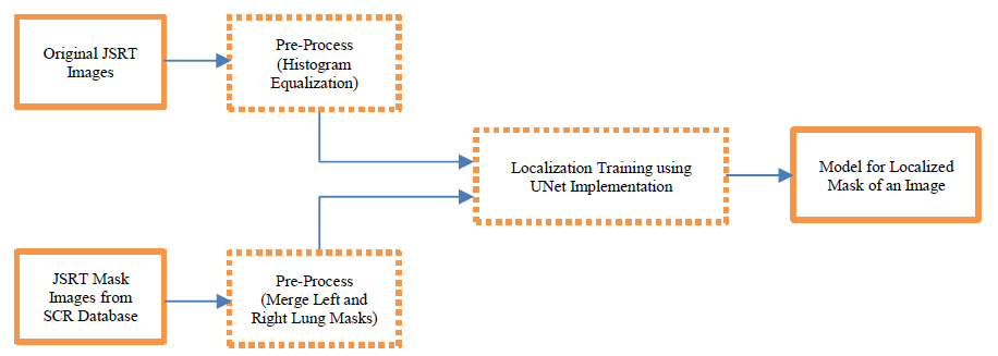
LeDNet: Localization-enabled Deep Neural Network for Multi-Label Radiography Image Classification
Lalit Pant, Shubham Arora
Multi-label radiography image classification has long been a topic of interest in neural networks research. In this paper, we intend to classify such images using convolution neural networks with novel localization techniques. We will use the chest x-ray images to detect thoracic diseases for this purpose. For accurate diagnosis, it is crucial to train the network with good quality images. But many chest X-ray images have irrelevant external objects like distractions created by faulty scans, electronic devices scanned next to lung region, scans inadvertently capturing bodily air etc. To address these, we propose a combination of localization and deep learning algorithms called LeDNet to predict thoracic diseases with higher accuracy. We identify and extract the lung region masks from chest x-ray images through localization. These masks are superimposed on the original X-ray images to create the mask overlay images. DenseNet-121 classification models are then used for feature selection to retrieve features of the entire chest X-ray images and the localized mask overlay images. These features are then used to predict disease classification. Our experiments involve comparing classification results obtained with original CheXpert images and mask overlay images. The comparison is demonstrated through accuracy and loss curve analyses.
Read more7/8/2024

0
Computer-Aided Diagnosis of Thoracic Diseases in Chest X-rays using hybrid CNN-Transformer Architecture
Sonit Singh
Medical imaging has been used for diagnosis of various conditions, making it one of the most powerful resources for effective patient care. Due to widespread availability, low cost, and low radiation, chest X-ray is one of the most sought after radiology examination for the diagnosis of various thoracic diseases. Due to advancements in medical imaging technologies and increasing patient load, current radiology workflow faces various challenges including increasing backlogs, working long hours, and increase in diagnostic errors. An automated computer-aided diagnosis system that can interpret chest X-rays to augment radiologists by providing actionable insights has potential to provide second opinion to radiologists, highlight relevant regions in the image, in turn expediting clinical workflow, reducing diagnostic errors, and improving patient care. In this study, we applied a novel architecture augmenting the DenseNet121 Convolutional Neural Network (CNN) with multi-head self-attention mechanism using transformer, namely SA-DenseNet121, that can identify multiple thoracic diseases in chest X-rays. We conducted experiments on four of the largest chest X-ray datasets, namely, ChestX-ray14, CheXpert, MIMIC-CXR-JPG, and IU-CXR. Experimental results in terms of area under the receiver operating characteristics (AUC-ROC) shows that augmenting CNN with self-attention has potential in diagnosing different thoracic diseases from chest X-rays. The proposed methodology has the potential to support the reading workflow, improve efficiency, and reduce diagnostic errors.
Read more4/22/2024