Leveraging the Mahalanobis Distance to enhance Unsupervised Brain MRI Anomaly Detection

0

Sign in to get full access
Overview
- This paper explores using the Mahalanobis distance, a statistical measure of distance, to enhance unsupervised anomaly detection in brain MRI scans.
- The authors propose a novel framework that leverages the Mahalanobis distance to identify anomalies in MRI images without the need for labeled training data.
- The approach aims to improve upon existing unsupervised anomaly detection methods, which can struggle with complex medical imaging data.
Plain English Explanation
The Mahalanobis distance is a way of measuring how different something is from the "typical" or "average" example. In this paper, the researchers use the Mahalanobis distance to identify abnormal or "anomalous" regions in brain MRI scans.
Typically, detecting abnormalities in medical images requires having a large dataset of labeled examples, which can be time-consuming and expensive to obtain. The key innovation in this work is that it uses an unsupervised approach, meaning the system can learn to detect anomalies without needing a pre-labeled dataset.
The core idea is to train a machine learning model to learn the "normal" patterns in healthy brain MRI scans. Then, when presented with a new scan, the model can use the Mahalanobis distance to identify regions that are significantly different from the learned "normal" patterns. These regions are flagged as potential anomalies or abnormalities.
By leveraging the Mahalanobis distance, the researchers aim to improve the performance of unsupervised anomaly detection in brain MRI scans, which could have important applications in medical diagnosis and disease monitoring.
Technical Explanation
The paper proposes a novel framework for unsupervised anomaly detection in brain MRI scans that leverages the Mahalanobis distance. The key steps are:
-
Feature Extraction: The researchers use a pre-trained ResNet model to extract visual features from the brain MRI scans.
-
Covariance Estimation: They estimate the covariance matrix of the extracted features from the "normal" (i.e., healthy) brain MRI scans.
-
Mahalanobis Distance Computation: For each voxel (3D pixel) in a new brain MRI scan, they compute the Mahalanobis distance from the "normal" covariance distribution.
-
Anomaly Scoring: Voxels with a high Mahalanobis distance are considered more anomalous and are assigned a higher anomaly score.
-
Anomaly Segmentation: The researchers use a GLAD-based approach to segment the anomalous regions in the MRI scan.
The authors evaluate their framework on several public brain MRI datasets and compare its performance to state-of-the-art unsupervised anomaly detection methods, such as UMAD and TAUAD. Their results demonstrate that the Mahalanobis distance-based approach outperforms these existing techniques, particularly in terms of accurate localization of anomalies.
Critical Analysis
The paper presents a well-designed and thorough evaluation of the proposed Mahalanobis distance-based anomaly detection framework. The authors acknowledge several limitations and areas for future research:
- The performance of the method may be sensitive to the choice of feature extraction model and hyperparameter tuning.
- The framework is currently evaluated on a limited set of public brain MRI datasets, and its generalization to a wider range of medical imaging data is not yet established.
- The interpretability of the Mahalanobis distance-based anomaly scores could be improved, which may be important for potential clinical applications.
Additionally, one could question the reliance on a pre-trained ResNet model for feature extraction, as this may introduce biases or limitations in the learned representations. Exploring end-to-end training approaches or using more specialized medical imaging architectures could be an area for further investigation.
Conclusion
This paper presents a novel unsupervised anomaly detection framework for brain MRI scans that leverages the Mahalanobis distance. By exploiting the statistical properties of "normal" brain MRI features, the proposed method demonstrates improved performance over existing unsupervised techniques, particularly in terms of localizing anomalous regions.
The work has the potential to advance the field of medical image analysis by enabling more accurate and efficient detection of abnormalities in brain MRI scans, which could aid in early disease diagnosis and monitoring. Further research is needed to explore the broader applicability of the Mahalanobis distance-based approach and address the identified limitations.
This summary was produced with help from an AI and may contain inaccuracies - check out the links to read the original source documents!
Related Papers
0
Leveraging the Mahalanobis Distance to enhance Unsupervised Brain MRI Anomaly Detection
Finn Behrendt, Debayan Bhattacharya, Robin Mieling, Lennart Maack, Julia Kruger, Roland Opfer, Alexander Schlaefer
Unsupervised Anomaly Detection (UAD) methods rely on healthy data distributions to identify anomalies as outliers. In brain MRI, a common approach is reconstruction-based UAD, where generative models reconstruct healthy brain MRIs, and anomalies are detected as deviations between input and reconstruction. However, this method is sensitive to imperfect reconstructions, leading to false positives that impede the segmentation. To address this limitation, we construct multiple reconstructions with probabilistic diffusion models. We then analyze the resulting distribution of these reconstructions using the Mahalanobis distance to identify anomalies as outliers. By leveraging information about normal variations and covariance of individual pixels within this distribution, we effectively refine anomaly scoring, leading to improved segmentation. Our experimental results demonstrate substantial performance improvements across various data sets. Specifically, compared to relying solely on single reconstructions, our approach achieves relative improvements of 15.9%, 35.4%, 48.0%, and 4.7% in terms of AUPRC for the BRATS21, ATLAS, MSLUB and WMH data sets, respectively.
Read more7/18/2024
0
Ensembled Cold-Diffusion Restorations for Unsupervised Anomaly Detection
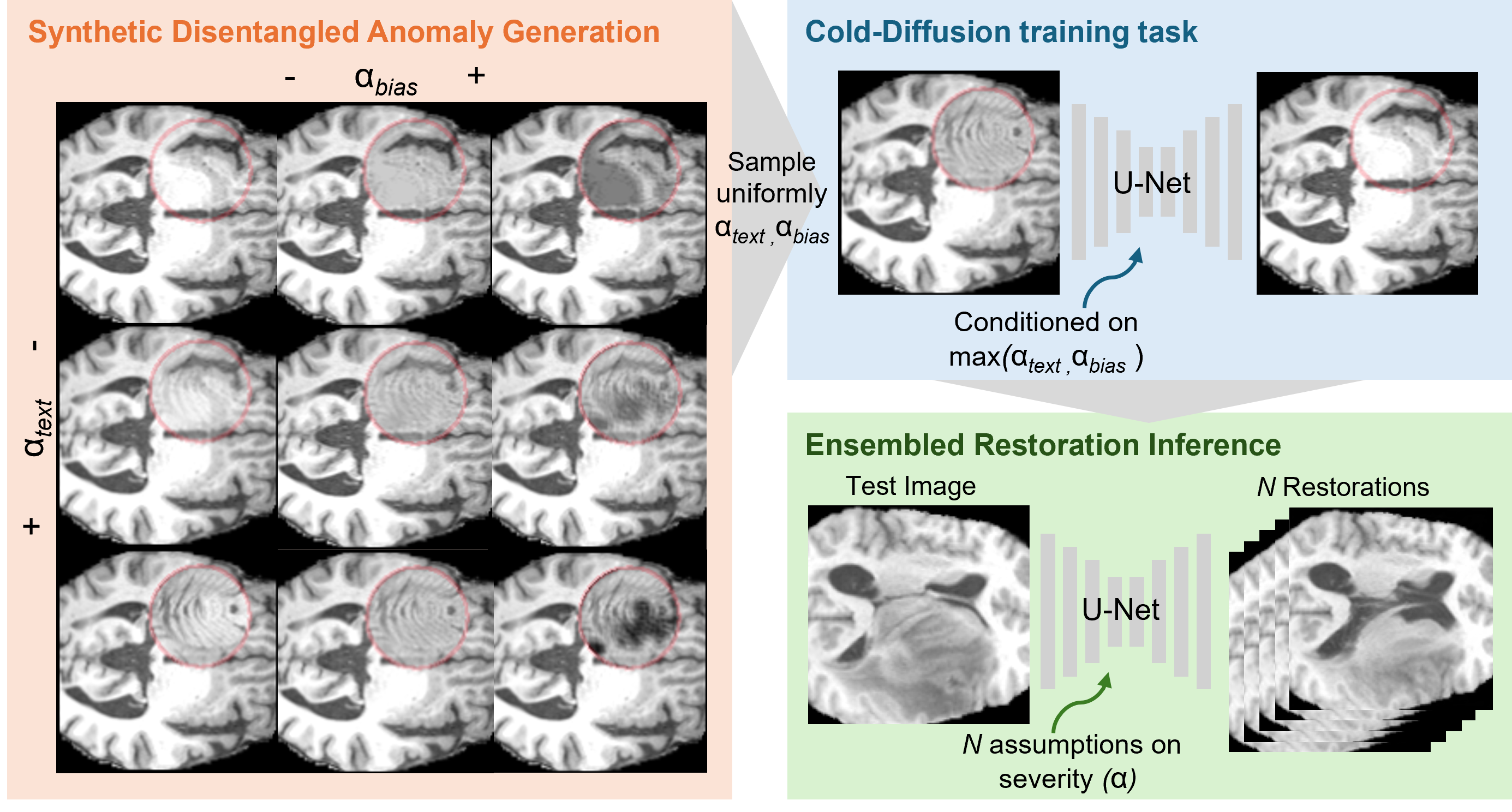
Sergio Naval Marimont, Vasilis Siomos, Matthew Baugh, Christos Tzelepis, Bernhard Kainz, Giacomo Tarroni
Unsupervised Anomaly Detection (UAD) methods aim to identify anomalies in test samples comparing them with a normative distribution learned from a dataset known to be anomaly-free. Approaches based on generative models offer interpretability by generating anomaly-free versions of test images, but are typically unable to identify subtle anomalies. Alternatively, approaches using feature modelling or self-supervised methods, such as the ones relying on synthetically generated anomalies, do not provide out-of-the-box interpretability. In this work, we present a novel method that combines the strengths of both strategies: a generative cold-diffusion pipeline (i.e., a diffusion-like pipeline which uses corruptions not based on noise) that is trained with the objective of turning synthetically-corrupted images back to their normal, original appearance. To support our pipeline we introduce a novel synthetic anomaly generation procedure, called DAG, and a novel anomaly score which ensembles restorations conditioned with different degrees of abnormality. Our method surpasses the prior state-of-the art for unsupervised anomaly detection in three different Brain MRI datasets.
Read more7/10/2024
🤿
0
Deep Learning-based Unsupervised Domain Adaptation via a Unified Model for Prostate Lesion Detection Using Multisite Bi-parametric MRI Datasets
Hao Li, Han Liu, Heinrich von Busch, Robert Grimm, Henkjan Huisman, Angela Tong, David Winkel, Tobias Penzkofer, Ivan Shabunin, Moon Hyung Choi, Qingsong Yang, Dieter Szolar, Steven Shea, Fergus Coakley, Mukesh Harisinghani, Ipek Oguz, Dorin Comaniciu, Ali Kamen, Bin Lou
Our hypothesis is that UDA using diffusion-weighted images, generated with a unified model, offers a promising and reliable strategy for enhancing the performance of supervised learning models in multi-site prostate lesion detection, especially when various b-values are present. This retrospective study included data from 5,150 patients (14,191 samples) collected across nine different imaging centers. A novel UDA method using a unified generative model was developed for multi-site PCa detection. This method translates diffusion-weighted imaging (DWI) acquisitions, including apparent diffusion coefficient (ADC) and individual DW images acquired using various b-values, to align with the style of images acquired using b-values recommended by Prostate Imaging Reporting and Data System (PI-RADS) guidelines. The generated ADC and DW images replace the original images for PCa detection. An independent set of 1,692 test cases (2,393 samples) was used for evaluation. The area under the receiver operating characteristic curve (AUC) was used as the primary metric, and statistical analysis was performed via bootstrapping. For all test cases, the AUC values for baseline SL and UDA methods were 0.73 and 0.79 (p=3, and 0.77 and 0.80 (p=4 PCa lesions. In the 361 test cases under the most unfavorable image acquisition setting, the AUC values for baseline SL and UDA were 0.49 and 0.76 (p=3, and 0.50 and 0.77 (p=4 PCa lesions. The results indicate the proposed UDA with generated images improved the performance of SL methods in multi-site PCa lesion detection across datasets with various b values, especially for images acquired with significant deviations from the PI-RADS recommended DWI protocol (e.g. with an extremely high b-value).
Read more8/12/2024

0
Rethinking Medical Anomaly Detection in Brain MRI: An Image Quality Assessment Perspective
Zixuan Pan, Jun Xia, Zheyu Yan, Guoyue Xu, Yawen Wu, Zhenge Jia, Jianxu Chen, Yiyu Shi
Reconstruction-based methods, particularly those leveraging autoencoders, have been widely adopted to perform anomaly detection in brain MRI. While most existing works try to improve detection accuracy by proposing new model structures or algorithms, we tackle the problem through image quality assessment, an underexplored perspective in the field. We propose a fusion quality loss function that combines Structural Similarity Index Measure loss with l1 loss, offering a more comprehensive evaluation of reconstruction quality. Additionally, we introduce a data pre-processing strategy that enhances the average intensity ratio (AIR) between normal and abnormal regions, further improving the distinction of anomalies. By fusing the aforementioned two methods, we devise the image quality assessment (IQA) approach. The proposed IQA approach achieves significant improvements (>10%) in terms of Dice coefficient (DICE) and Area Under the Precision-Recall Curve (AUPRC) on the BraTS21 (T2, FLAIR) and MSULB datasets when compared with state-of-the-art methods. These results highlight the importance of invoking the comprehensive image quality assessment in medical anomaly detection and provide a new perspective for future research in this field.
Read more8/16/2024