Monocular pose estimation of articulated surgical instruments in open surgery

0
Sign in to get full access
Overview
- Presents a method for estimating the 3D pose of articulated surgical instruments from monocular camera images during open surgery.
- Leverages a neural network architecture to extract features from the image and predict the 6D pose of the instrument.
- Demonstrates the approach on a dataset of surgical instrument images, showing improved accuracy over existing techniques.
Plain English Explanation
This research paper describes a new way to track the position and orientation of surgical instruments used in open surgery. The key idea is to use a deep learning model that can analyze a single camera image and figure out the 3D pose (position and orientation) of the surgical instrument, even if it has multiple moving parts.
This is useful because it allows surgeons and surgical robots to better understand the location and movement of instruments during procedures, which can help improve precision and safety. Existing methods for tracking surgical instruments often require special sensors or markers, which can be cumbersome or impractical in real-world surgical settings. The approach presented in this paper instead relies only on standard camera images, making it potentially more practical and scalable.
The researchers tested their method on a dataset of images showing surgical instruments, and found that it was able to estimate the 3D pose of the instruments more accurately than previous techniques. This suggests the model is effectively learning to extract the relevant visual features from the images to infer the instrument's position and orientation.
Technical Explanation
The paper proposes a neural network-based approach for monocular 3D pose estimation of articulated surgical instruments. The key components include:
- A deep neural network architecture that takes a single RGB camera image as input and outputs the 6D pose (3D position and 3D orientation) of the surgical instrument.
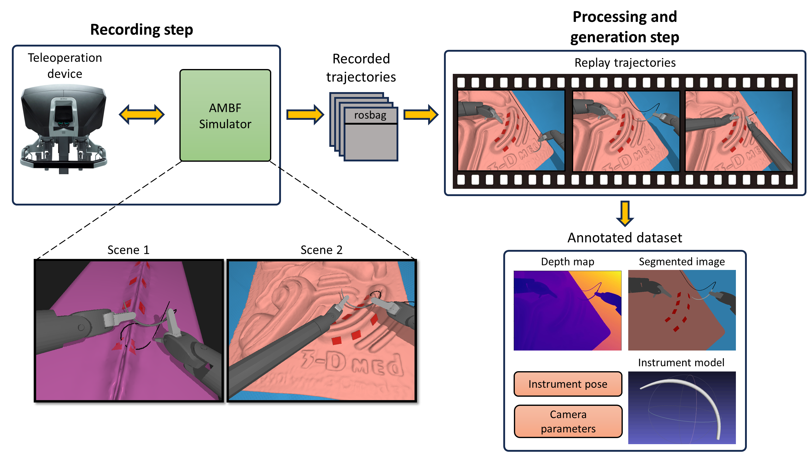
- A novel data generation pipeline to create a large and diverse training dataset of synthetic instrument images with ground truth pose labels.
- Head pose estimation and surgical tool detection modules to further improve pose estimation accuracy.
The model is evaluated on a dataset of real surgical instrument images, demonstrating improved 6D pose estimation performance compared to prior state-of-the-art methods.
Critical Analysis
The paper presents a comprehensive technical approach to a challenging problem in computer vision for surgical applications. The use of synthetic data generation and integration of additional modules like head pose estimation are thoughtful design choices that likely contribute to the strong empirical results.
That said, the paper does not deeply explore the limitations of the proposed method. For example, it is unclear how the system would perform on more diverse and cluttered surgical scenes, or how sensitive it is to changes in lighting, camera viewpoint, or instrument appearance. Additionally, the evaluation is limited to a single dataset, and further testing on more diverse surgical settings would be valuable.
Overall, this research represents an important step forward in enabling robust and practical 3D pose tracking of articulated surgical instruments. However, additional work is likely needed to fully validate the approach and understand its real-world applicability in the complex and dynamic environments of actual surgical procedures.
Conclusion
This paper presents a novel deep learning-based method for estimating the 3D pose of articulated surgical instruments from monocular camera images. By leveraging a tailored neural network architecture and a synthetic data generation pipeline, the approach demonstrates improved pose estimation accuracy over prior techniques.
The ability to track the position and orientation of surgical instruments without specialized sensors or markers could enable more advanced computer-assisted surgical systems and enhanced situational awareness for surgeons. While additional research is needed to fully validate the method's robustness and generalizability, this work represents an important contribution to the field of computer vision for surgical applications.
This summary was produced with help from an AI and may contain inaccuracies - check out the links to read the original source documents!
Related Papers
0
Monocular pose estimation of articulated surgical instruments in open surgery
Robert Spektor, Tom Friedman, Itay Or, Gil Bolotin, Shlomi Laufer
This work presents a novel approach to monocular 6D pose estimation of surgical instruments in open surgery, addressing challenges such as object articulations, symmetries, occlusions, and lack of annotated real-world data. The method leverages synthetic data generation and domain adaptation techniques to overcome these obstacles. The proposed approach consists of three main components: (1) synthetic data generation using 3D modeling of surgical tools with articulation rigging and physically-based rendering; (2) a tailored pose estimation framework combining object detection with pose estimation and a hybrid geometric fusion strategy; and (3) a training strategy that utilizes both synthetic and real unannotated data, employing domain adaptation on real video data using automatically generated pseudo-labels. Evaluations conducted on videos of open surgery demonstrate the good performance and real-world applicability of the proposed method, highlighting its potential for integration into medical augmented reality and robotic systems. The approach eliminates the need for extensive manual annotation of real surgical data.
Read more7/18/2024
0
Realistic Data Generation for 6D Pose Estimation of Surgical Instruments
Juan Antonio Barragan, Jintan Zhang, Haoying Zhou, Adnan Munawar, Peter Kazanzides
Automation in surgical robotics has the potential to improve patient safety and surgical efficiency, but it is difficult to achieve due to the need for robust perception algorithms. In particular, 6D pose estimation of surgical instruments is critical to enable the automatic execution of surgical maneuvers based on visual feedback. In recent years, supervised deep learning algorithms have shown increasingly better performance at 6D pose estimation tasks; yet, their success depends on the availability of large amounts of annotated data. In household and industrial settings, synthetic data, generated with 3D computer graphics software, has been shown as an alternative to minimize annotation costs of 6D pose datasets. However, this strategy does not translate well to surgical domains as commercial graphics software have limited tools to generate images depicting realistic instrument-tissue interactions. To address these limitations, we propose an improved simulation environment for surgical robotics that enables the automatic generation of large and diverse datasets for 6D pose estimation of surgical instruments. Among the improvements, we developed an automated data generation pipeline and an improved surgical scene. To show the applicability of our system, we generated a dataset of 7.5k images with pose annotations of a surgical needle that was used to evaluate a state-of-the-art pose estimation network. The trained model obtained a mean translational error of 2.59mm on a challenging dataset that presented varying levels of occlusion. These results highlight our pipeline's success in training and evaluating novel vision algorithms for surgical robotics applications.
Read more6/12/2024

0
Advancing 6-DoF Instrument Pose Estimation in Variable X-Ray Imaging Geometries
Christiaan G. A. Viviers, Lena Filatova, Maurice Termeer, Peter H. N. de With, Fons van der Sommen
Accurate 6-DoF pose estimation of surgical instruments during minimally invasive surgeries can substantially improve treatment strategies and eventual surgical outcome. Existing deep learning methods have achieved accurate results, but they require custom approaches for each object and laborious setup and training environments often stretching to extensive simulations, whilst lacking real-time computation. We propose a general-purpose approach of data acquisition for 6-DoF pose estimation tasks in X-ray systems, a novel and general purpose YOLOv5-6D pose architecture for accurate and fast object pose estimation and a complete method for surgical screw pose estimation under acquisition geometry consideration from a monocular cone-beam X-ray image. The proposed YOLOv5-6D pose model achieves competitive results on public benchmarks whilst being considerably faster at 42 FPS on GPU. In addition, the method generalizes across varying X-ray acquisition geometry and semantic image complexity to enable accurate pose estimation over different domains. Finally, the proposed approach is tested for bone-screw pose estimation for computer-aided guidance during spine surgeries. The model achieves a 92.41% by the 0.1 ADD-S metric, demonstrating a promising approach for enhancing surgical precision and patient outcomes. The code for YOLOv5-6D is publicly available at https://github.com/cviviers/YOLOv5-6D-Pose
Read more5/21/2024

0
Head Pose Estimation and 3D Neural Surface Reconstruction via Monocular Camera in situ for Navigation and Safe Insertion into Natural Openings
Ruijie Tang, Beilei Cui, Hongliang Ren
As the significance of simulation in medical care and intervention continues to grow, it is anticipated that a simplified and low-cost platform can be set up to execute personalized diagnoses and treatments. 3D Slicer can not only perform medical image analysis and visualization but can also provide surgical navigation and surgical planning functions. In this paper, we have chosen 3D Slicer as our base platform and monocular cameras are used as sensors. Then, We used the neural radiance fields (NeRF) algorithm to complete the 3D model reconstruction of the human head. We compared the accuracy of the NeRF algorithm in generating 3D human head scenes and utilized the MarchingCube algorithm to generate corresponding 3D mesh models. The individual's head pose, obtained through single-camera vision, is transmitted in real-time to the scene created within 3D Slicer. The demonstrations presented in this paper include real-time synchronization of transformations between the human head model in the 3D Slicer scene and the detected head posture. Additionally, we tested a scene where a tool, marked with an ArUco Maker tracked by a single camera, synchronously points to the real-time transformation of the head posture. These demos indicate that our methodology can provide a feasible real-time simulation platform for nasopharyngeal swab collection or intubation.
Read more6/21/2024