Neurovascular Segmentation in sOCT with Deep Learning and Synthetic Training Data

0
Sign in to get full access
Overview
- This paper explores the use of deep learning and synthetic training data for neurovascular segmentation in swept-source optical coherence tomography (sOCT) imaging.
- The researchers developed a novel deep learning model and leveraged synthetic training data to address the challenges of limited real-world datasets and high variability in vascular structures.
- The proposed approach demonstrates significant improvements in segmentation accuracy compared to traditional methods, paving the way for more robust and automated vascular analysis in biomedical imaging applications.
Plain English Explanation
Biomedical imaging techniques, such as sOCT, are widely used to study the intricate network of blood vessels, or vasculature, in the human body. Accurately segmenting and identifying these vascular structures is crucial for various medical applications, including stroke diagnosis and cardiovascular disease monitoring.
The authors of this paper tackled the challenge of automating the process of vascular segmentation using deep learning, a powerful artificial intelligence technique. They developed a new deep learning model, specifically designed for the task of neurovascular segmentation in sOCT images.
One of the key innovations in this research is the use of synthetic training data. Real-world sOCT datasets can be limited in size and may not capture the full diversity of vascular structures. To address this, the researchers created realistic synthetic sOCT images with simulated vascular networks. By training their deep learning model on this synthetic data, along with a smaller set of real-world images, they were able to achieve significantly improved segmentation accuracy compared to traditional methods.
The approach described in this paper represents an important step forward in automating vascular analysis and could have far-reaching implications for various medical applications, such as early disease detection and personalized healthcare.
Technical Explanation
The paper proposes a deep learning-based approach for neurovascular segmentation in sOCT imaging, leveraging synthetic training data to address the challenges of limited real-world datasets and high variability in vascular structures.
The researchers developed a novel deep learning model, referred to as the Synthetic Augmented Neurovascular Segmentation (SANS) network. SANS is a convolutional neural network (CNN) architecture designed specifically for the task of vascular segmentation in sOCT images.
To overcome the limitations of real-world sOCT datasets, the authors created a large-scale synthetic dataset of sOCT images with realistic vascular structures. They used a physics-based rendering engine to simulate the optical properties of tissue and vasculature, generating high-quality synthetic sOCT volumes. These synthetic images, along with a smaller set of real-world sOCT data, were used to train the SANS network.
The experiments conducted in the paper demonstrate that the SANS network, trained on the combination of synthetic and real-world data, outperforms traditional segmentation methods, as well as deep learning models trained on real-world data alone. The researchers report significant improvements in segmentation accuracy, particularly in the ability to capture fine-grained vascular structures that are often challenging to detect in sOCT images.
Critical Analysis
The paper presents a comprehensive and well-designed study, addressing the important challenge of automating vascular segmentation in biomedical imaging. The use of synthetic training data is a notable strength of the proposed approach, as it allows the deep learning model to learn from a diverse set of vascular structures that may not be readily available in real-world datasets.
However, the authors acknowledge that the synthetic data generation process may not fully capture all the complexities and variability present in real-world sOCT images. While the experiments demonstrate the effectiveness of the SANS network, further research is needed to investigate the model's generalization capabilities across different imaging modalities and patient populations.
Additionally, the paper does not provide a detailed analysis of the computational complexity and inference time of the SANS network, which are important practical considerations for real-world deployment in clinical settings. The authors could also explore the potential for incorporating active learning or other techniques to further improve the model's performance with limited real-world data.
Conclusion
This paper presents a significant contribution to the field of biomedical image analysis, demonstrating the potential of deep learning and synthetic training data for robust and automated vascular segmentation in sOCT imaging. The proposed SANS network, trained on a combination of synthetic and real-world data, outperforms traditional segmentation methods and sets a new benchmark for neurovascular segmentation.
The findings of this research have important implications for various medical applications, such as early disease detection, personalized healthcare, and the development of advanced diagnostic and monitoring tools. By automating the analysis of vascular structures, the approach described in this paper could lead to more efficient and accurate clinical decision-making, ultimately improving patient outcomes.
This summary was produced with help from an AI and may contain inaccuracies - check out the links to read the original source documents!
Related Papers
0
Neurovascular Segmentation in sOCT with Deep Learning and Synthetic Training Data
Etienne Chollet, Yael Balbastre, Chiara Mauri, Caroline Magnain, Bruce Fischl, Hui Wang
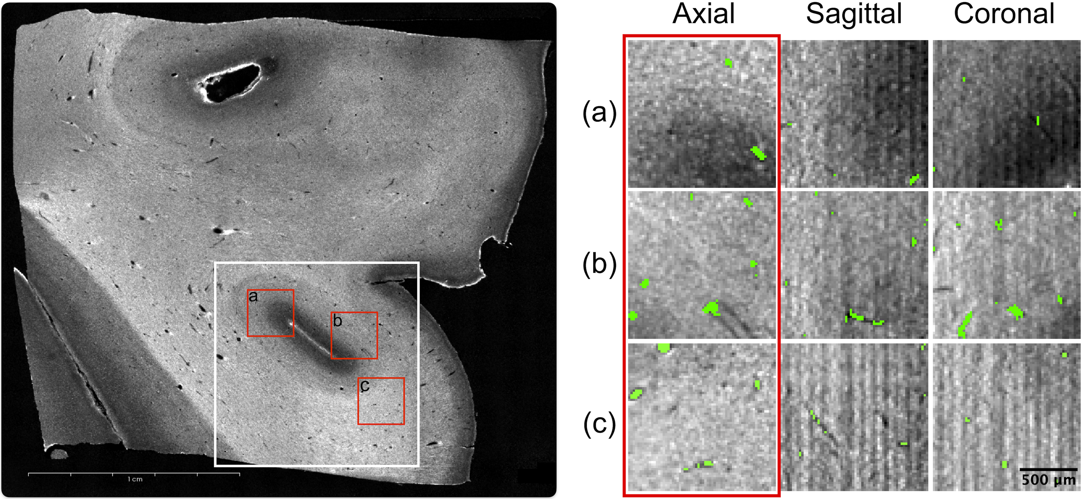
Microvascular anatomy is known to be involved in various neurological disorders. However, understanding these disorders is hindered by the lack of imaging modalities capable of capturing the comprehensive three-dimensional vascular network structure at microscopic resolution. With a lateral resolution of $<=$20 {textmu}m and ability to reconstruct large tissue blocks up to tens of cubic centimeters, serial-section optical coherence tomography (sOCT) is well suited for this task. This method uses intrinsic optical properties to visualize the vessels and therefore does not possess a specific contrast, which complicates the extraction of accurate vascular models. The performance of traditional vessel segmentation methods is heavily degraded in the presence of substantial noise and imaging artifacts and is sensitive to domain shifts, while convolutional neural networks (CNNs) require extensive labeled data and are also sensitive the precise intensity characteristics of the data that they are trained on. Building on the emerging field of synthesis-based training, this study demonstrates a synthesis engine for neurovascular segmentation in sOCT images. Characterized by minimal priors and high variance sampling, our highly generalizable method tested on five distinct sOCT acquisitions eliminates the need for manual annotations while attaining human-level precision. Our approach comprises two phases: label synthesis and label-to-image transformation. We demonstrate the efficacy of the former by comparing it to several more realistic sets of training labels, and the latter by an ablation study of synthetic noise and artifact models.
Read more7/2/2024

0
A label-free and data-free training strategy for vasculature segmentation in serial sectioning OCT data
Etienne Chollet, Yael Balbastre, Caroline Magnain, Bruce Fischl, Hui Wang
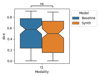
Serial sectioning Optical Coherence Tomography (sOCT) is a high-throughput, label free microscopic imaging technique that is becoming increasingly popular to study post-mortem neurovasculature. Quantitative analysis of the vasculature requires highly accurate segmentation; however, sOCT has low signal-to-noise-ratio and displays a wide range of contrasts and artifacts that depend on acquisition parameters. Furthermore, labeled data is scarce and extremely time consuming to generate. Here, we leverage synthetic datasets of vessels to train a deep learning segmentation model. We construct the vessels with semi-realistic splines that simulate the vascular geometry and compare our model with realistic vascular labels generated by constrained constructive optimization. Both approaches yield similar Dice scores, although with very different false positive and false negative rates. This method addresses the complexity inherent in OCT images and paves the way for more accurate and efficient analysis of neurovascular structures.
Read more5/24/2024
0
Synthetic Data for Robust Stroke Segmentation
Liam Chalcroft, Ioannis Pappas, Cathy J. Price, John Ashburner
Deep learning-based semantic segmentation in neuroimaging currently requires high-resolution scans and extensive annotated datasets, posing significant barriers to clinical applicability. We present a novel synthetic framework for the task of lesion segmentation, extending the capabilities of the established SynthSeg approach to accommodate large heterogeneous pathologies with lesion-specific augmentation strategies. Our method trains deep learning models, demonstrated here with the UNet architecture, using label maps derived from healthy and stroke datasets, facilitating the segmentation of both healthy tissue and pathological lesions without sequence-specific training data. Evaluated against in-domain and out-of-domain (OOD) datasets, our framework demonstrates robust performance, rivaling current methods within the training domain and significantly outperforming them on OOD data. This contribution holds promise for advancing medical imaging analysis in clinical settings, especially for stroke pathology, by enabling reliable segmentation across varied imaging sequences with reduced dependency on large annotated corpora. Code and weights available at https://github.com/liamchalcroft/SynthStroke.
Read more4/3/2024
🤿
0
Automating Vessel Segmentation in the Heart and Brain: A Trend to Develop Multi-Modality and Label-Efficient Deep Learning Techniques
Nazik Elsayed, Yousuf Babiker M. Osman, Cheng Li, Jiong Zhang, Shanshan Wang
Cardio-cerebrovascular diseases are the leading causes of mortality worldwide, whose accurate blood vessel segmentation is significant for both scientific research and clinical usage. However, segmenting cardio-cerebrovascular structures from medical images is very challenging due to the presence of thin or blurred vascular shapes, imbalanced distribution of vessel and non-vessel pixels, and interference from imaging artifacts. These difficulties make manual or semi-manual segmentation methods highly time-consuming, labor-intensive, and prone to errors with interobserver variability, where different experts may produce different segmentations from a variety of modalities. Consequently, there is a growing interest in developing automated algorithms. This paper provides an up-to-date survey of deep learning techniques, for cardio-cerebrovascular segmentation. It analyzes the research landscape, surveys recent approaches, and discusses challenges such as the scarcity of accurately annotated data and variability. This paper also illustrates the urgent needs for developing multi-modality label-efficient deep learning techniques. To the best of our knowledge, this paper is the first comprehensive survey of deep learning approaches that effectively segment vessels in both the heart and brain. It aims to advance automated segmentation techniques for cardio-cerebrovascular diseases, benefiting researchers and healthcare professionals.
Read more4/3/2024