NMGrad: Advancing Histopathological Bladder Cancer Grading with Weakly Supervised Deep Learning

0

Sign in to get full access
Overview
- This paper proposes a weakly supervised deep learning framework called NMGrad to improve the accuracy of histopathological bladder cancer grading.
- Bladder cancer grading is an important task, but current approaches rely on manual, subjective evaluation by pathologists, which can be inconsistent.
- The NMGrad framework aims to leverage weakly labeled data, where only the overall cancer grade is known, to train a model that can provide more accurate and consistent grading of individual tissue samples.
Plain English Explanation
The research paper introduces a new machine learning technique called NMGrad that can help doctors more accurately diagnose and grade bladder cancer from tissue samples. Currently, doctors visually inspect tissue samples under a microscope to determine the cancer grade, but this process can be subjective and inconsistent between different doctors.
The NMGrad approach takes advantage of "weakly labeled" data, where the overall cancer grade is known but the grade of individual tissue samples is not. By training a deep learning model on this type of data, the researchers were able to develop a system that can more accurately grade individual tissue samples, rather than relying solely on a doctor's visual inspection. This is an important advance, as more accurate grading can help doctors provide better treatment recommendations for bladder cancer patients.
The key idea behind NMGrad is to leverage the known overall cancer grade to guide the model in learning to recognize the visual patterns associated with different cancer grades, even when the grade of each individual tissue sample is not provided. This "weakly supervised" approach allows the model to be trained on a larger and more diverse dataset compared to traditional approaches that require each tissue sample to be manually graded.
Technical Explanation
The NMGrad framework proposed in this paper aims to address the challenge of inconsistent and subjective bladder cancer grading by pathologists. The researchers developed a weakly supervised deep learning approach that can learn to grade individual tissue samples from data where only the overall cancer grade is known.
The core of the NMGrad approach is a convolutional neural network (CNN) that takes a tissue sample image as input and predicts the corresponding cancer grade. To train this model, the researchers leveraged a "weakly supervised" learning setup, where the training data consists of tissue sample images and their associated overall cancer grade, but the individual grade of each tissue sample is not provided.
The key innovation of NMGrad is a novel loss function that guides the CNN model to learn visual patterns associated with different cancer grades, even without direct supervision of the individual tissue sample grades. This loss function combines a standard classification loss with a "gradient-based consistency" term that encourages the model's predictions to be consistent with the known overall cancer grade.
Through extensive experiments on a large bladder cancer histopathology dataset, the researchers demonstrated that the NMGrad framework outperforms previous state-of-the-art approaches for bladder cancer grading. The NMGrad model achieved significantly higher accuracy in predicting the individual tissue sample grades compared to models trained with traditional fully-supervised techniques.
Critical Analysis
The NMGrad framework represents an important advance in the field of computational pathology and automated cancer diagnosis. By leveraging weakly supervised learning, the researchers were able to develop a more scalable and practical approach compared to previous methods that required time-consuming manual annotation of individual tissue samples.
However, the paper does not address several potential limitations and areas for further research. For example, the performance of the NMGrad model may be sensitive to the quality and diversity of the training data, and it is unclear how well the approach would generalize to datasets from different institutions or with different imaging modalities.
Additionally, the paper does not provide a detailed analysis of the types of visual patterns the NMGrad model learns to recognize, nor does it offer insights into the model's decision-making process. Incorporating more interpretable AI techniques could help build trust in the model's predictions and facilitate its adoption by healthcare professionals.
Further research is also needed to explore the potential clinical impact of the NMGrad framework, such as its ability to improve patient outcomes or streamline the diagnostic workflow. Integrating the NMGrad model into a comprehensive AI-powered cancer diagnosis system could be a promising direction for future work.
Conclusion
The NMGrad framework presented in this paper represents a significant advance in the field of computational pathology and automated cancer diagnosis. By leveraging weakly supervised deep learning, the researchers were able to develop a model that can more accurately grade individual bladder cancer tissue samples, overcoming the limitations of subjective and inconsistent manual grading by pathologists.
The NMGrad approach demonstrates the potential of advanced AI techniques to improve healthcare workflows and patient outcomes. While further research is needed to address the limitations and explore the clinical impact of this framework, the NMGrad model shows promise as a valuable tool to support more consistent and reliable bladder cancer diagnosis and treatment.
This summary was produced with help from an AI and may contain inaccuracies - check out the links to read the original source documents!
Related Papers
0
NMGrad: Advancing Histopathological Bladder Cancer Grading with Weakly Supervised Deep Learning
Saul Fuster, Umay Kiraz, Trygve Eftest{o}l, Emiel A. M. Janssen, Kjersti Engan
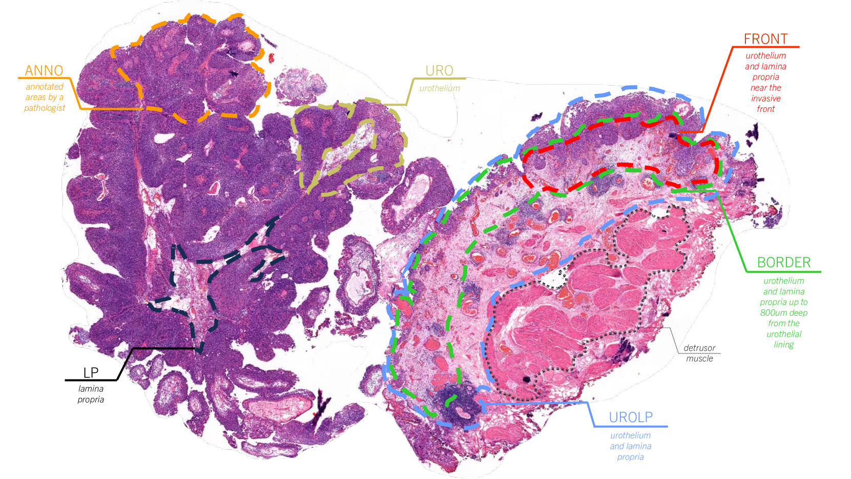
The most prevalent form of bladder cancer is urothelial carcinoma, characterized by a high recurrence rate and substantial lifetime treatment costs for patients. Grading is a prime factor for patient risk stratification, although it suffers from inconsistencies and variations among pathologists. Moreover, absence of annotations in medical imaging difficults training deep learning models. To address these challenges, we introduce a pipeline designed for bladder cancer grading using histological slides. First, it extracts urothelium tissue tiles at different magnification levels, employing a convolutional neural network for processing for feature extraction. Then, it engages in the slide-level prediction process. It employs a nested multiple instance learning approach with attention to predict the grade. To distinguish different levels of malignancy within specific regions of the slide, we include the origins of the tiles in our analysis. The attention scores at region level is shown to correlate with verified high-grade regions, giving some explainability to the model. Clinical evaluations demonstrate that our model consistently outperforms previous state-of-the-art methods.
Read more5/27/2024
0
Self-Contrastive Weakly Supervised Learning Framework for Prognostic Prediction Using Whole Slide Images
Saul Fuster, Farbod Khoraminia, Julio Silva-Rodr'iguez, Umay Kiraz, Geert J. L. H. van Leenders, Trygve Eftest{o}l, Valery Naranjo, Emiel A. M. Janssen, Tahlita C. M. Zuiverloon, Kjersti Engan
We present a pioneering investigation into the application of deep learning techniques to analyze histopathological images for addressing the substantial challenge of automated prognostic prediction. Prognostic prediction poses a unique challenge as the ground truth labels are inherently weak, and the model must anticipate future events that are not directly observable in the image. To address this challenge, we propose a novel three-part framework comprising of a convolutional network based tissue segmentation algorithm for region of interest delineation, a contrastive learning module for feature extraction, and a nested multiple instance learning classification module. Our study explores the significance of various regions of interest within the histopathological slides and exploits diverse learning scenarios. The pipeline is initially validated on artificially generated data and a simpler diagnostic task. Transitioning to prognostic prediction, tasks become more challenging. Employing bladder cancer as use case, our best models yield an AUC of 0.721 and 0.678 for recurrence and treatment outcome prediction respectively.
Read more5/27/2024

0
Predicting the risk of early-stage breast cancer recurrence using H&E-stained tissue images
Geongyu Lee, Joonho Lee, Tae-Yeong Kwak, Sun Woo Kim, Youngmee Kwon, Chungyeul Kim, Hyeyoon Chang
Accurate prediction of the likelihood of recurrence is important in the selection of postoperative treatment for patients with early-stage breast cancer. In this study, we investigated whether deep learning algorithms can predict patients' risk of recurrence by analyzing the pathology images of their cancer histology. A total of 125 hematoxylin and eosin stained breast cancer whole slide images labeled with the risk prediction via genomics assays were used, and we obtained sensitivity of 0.857, 0.746, and 0.529 for predicting low, intermediate, and high risk, and specificity of 0.816, 0.803, and 0.972. When compared to the expert pathologist's regional histology grade information, a Pearson's correlation coefficient of 0.61 was obtained. When we checked the model learned through these studies through the class activation map, we found that it actually considered tubule formation and mitotic rate when predicting different risk groups.
Read more6/12/2024
🖼️
0
An interpretable machine learning system for colorectal cancer diagnosis from pathology slides
Pedro C. Neto, Diana Montezuma, Sara P. Oliveira, Domingos Oliveira, Jo~ao Fraga, Ana Monteiro, Jo~ao Monteiro, Liliana Ribeiro, Sofia Gonc{c}alves, Stefan Reinhard, Inti Zlobec, Isabel M. Pinto, Jaime S. Cardoso
Considering the profound transformation affecting pathology practice, we aimed to develop a scalable artificial intelligence (AI) system to diagnose colorectal cancer from whole-slide images (WSI). For this, we propose a deep learning (DL) system that learns from weak labels, a sampling strategy that reduces the number of training samples by a factor of six without compromising performance, an approach to leverage a small subset of fully annotated samples, and a prototype with explainable predictions, active learning features and parallelisation. Noting some problems in the literature, this study is conducted with one of the largest WSI colorectal samples dataset with approximately 10,500 WSIs. Of these samples, 900 are testing samples. Furthermore, the robustness of the proposed method is assessed with two additional external datasets (TCGA and PAIP) and a dataset of samples collected directly from the proposed prototype. Our proposed method predicts, for the patch-based tiles, a class based on the severity of the dysplasia and uses that information to classify the whole slide. It is trained with an interpretable mixed-supervision scheme to leverage the domain knowledge introduced by pathologists through spatial annotations. The mixed-supervision scheme allowed for an intelligent sampling strategy effectively evaluated in several different scenarios without compromising the performance. On the internal dataset, the method shows an accuracy of 93.44% and a sensitivity between positive (low-grade and high-grade dysplasia) and non-neoplastic samples of 0.996. On the external test samples varied with TCGA being the most challenging dataset with an overall accuracy of 84.91% and a sensitivity of 0.996.
Read more5/2/2024