Reference-Free Multi-Modality Volume Registration of X-Ray Microscopy and Light-Sheet Fluorescence Microscopy

0
🤷
Sign in to get full access
Overview
- X-ray microscopy (XRM) and light-sheet fluorescence microscopy (LSFM) are emerging imaging techniques used in preclinical research on bone remodeling diseases
- These complementary methods offer micrometer-level resolution, providing a holistic view of bone microstructures
- Registering these independently acquired large-scale volumes is extremely challenging without reference points
Plain English Explanation
Researchers have developed two new imaging tools, X-ray microscopy (XRM) and light-sheet fluorescence microscopy (LSFM), that can capture detailed images of bone structure at the micrometer level. By combining the information from these two complementary techniques, scientists can get a more comprehensive understanding of how bones change over the course of different diseases, like osteoporosis.
However, aligning the large 3D data sets from these independent imaging methods is extremely difficult, especially when there are no reference points to use as landmarks. This paper presents a new two-stage process to quickly and accurately register the XRM and LSFM data, enabling researchers to precisely match up features like bone cells and tiny cavities (lacunae) across the different imaging modalities. This allows them to gain novel insights into how bone structure and function are affected by diseases that are common as people age.
Technical Explanation
The proposed pipeline has two main stages. In the first stage, the method extracts surface features from the XRM and LSFM data and uses two successive point cloud-based alignment techniques to achieve a coarse registration.
The second stage then fine-tunes this initial alignment using a modified cross-correlation method to ensure precise volumetric registration between the two imaging modalities. The authors also introduce a novel "residual similarity" metric to quantitatively assess the quality of the final alignment.
The results demonstrate a robust, gradual improvement in registration accuracy across the two stages of the pipeline. Ultimately, this allows the researchers to accurately match up key bone microstructures observed in the XRM and LSFM data, such as the lacunae (tiny cavities) and bone cells. This integrated view provides new opportunities to study diseases like osteoporosis, which are a significant burden on aging populations.
Critical Analysis
The paper presents a promising approach to overcoming the challenging problem of registering XRM and LSFM data, which are crucial for advancing our understanding of bone remodeling diseases. The authors' use of a two-stage process with point cloud-based coarse alignment followed by fine-tuning using cross-correlation seems well-designed to handle the complexities of aligning these large-scale, independently acquired volumes.
However, the paper does not address potential limitations or edge cases, such as how the method might perform with data sets that have significant differences in resolution, field of view, or signal-to-noise ratio between the XRM and LSFM modalities. Additionally, while the authors introduce a new "residual similarity" metric, they do not provide a thorough validation of its usefulness compared to other potential evaluation approaches.
Further research could explore the generalizability of this registration pipeline to other types of multimodal biomedical imaging data, as well as investigate ways to make the process even more robust and automated. Nonetheless, this work represents an important step forward in integrating complementary imaging techniques to gain a more holistic understanding of complex biological structures and diseases.
Conclusion
This paper presents a fast and effective two-stage pipeline for registering XRM and LSFM data, which are powerful imaging tools for studying bone remodeling at the micrometer scale. By aligning these complementary modalities, the method enables researchers to precisely match up key bone microstructures, such as lacunae and bone cells, across the different imaging techniques.
This integrated view can provide valuable new insights into diseases like osteoporosis, which are a significant burden on aging populations. While the paper does not address all potential limitations, it represents an important advance in overcoming the challenges of multimodal biomedical image registration, paving the way for more comprehensive, function-oriented analyses of complex biological systems.
This summary was produced with help from an AI and may contain inaccuracies - check out the links to read the original source documents!
Related Papers
🤷
0
Reference-Free Multi-Modality Volume Registration of X-Ray Microscopy and Light-Sheet Fluorescence Microscopy
Siyuan Mei, Fuxin Fan, Mareike Thies, Mingxuan Gu, Fabian Wagner, Oliver Aust, Ina Erceg, Zeynab Mirzaei, Georgiana Neag, Yipeng Sun, Yixing Huang, Andreas Maier
Recently, X-ray microscopy (XRM) and light-sheet fluorescence microscopy (LSFM) have emerged as two pivotal imaging tools in preclinical research on bone remodeling diseases, offering micrometer-level resolution. Integrating these complementary modalities provides a holistic view of bone microstructures, facilitating function-oriented volume analysis across different disease cycles. However, registering such independently acquired large-scale volumes is extremely challenging under real and reference-free scenarios. This paper presents a fast two-stage pipeline for volume registration of XRM and LSFM. The first stage extracts the surface features and employs two successive point cloud-based methods for coarse alignment. The second stage fine-tunes the initial alignment using a modified cross-correlation method, ensuring precise volumetric registration. Moreover, we propose residual similarity as a novel metric to assess the alignment of two complementary modalities. The results imply robust gradual improvement across the stages. In the end, all correlating microstructures, particularly lacunae in XRM and bone cells in LSFM, are precisely matched, enabling new insights into bone diseases like osteoporosis which are a substantial burden in aging societies.
Read more4/24/2024

0
Super-resolution of biomedical volumes with 2D supervision
Cheng Jiang, Alexander Gedeon, Yiwei Lyu, Eric Landgraf, Yufeng Zhang, Xinhai Hou, Akhil Kondepudi, Asadur Chowdury, Honglak Lee, Todd Hollon
Volumetric biomedical microscopy has the potential to increase the diagnostic information extracted from clinical tissue specimens and improve the diagnostic accuracy of both human pathologists and computational pathology models. Unfortunately, barriers to integrating 3-dimensional (3D) volumetric microscopy into clinical medicine include long imaging times, poor depth / z-axis resolution, and an insufficient amount of high-quality volumetric data. Leveraging the abundance of high-resolution 2D microscopy data, we introduce masked slice diffusion for super-resolution (MSDSR), which exploits the inherent equivalence in the data-generating distribution across all spatial dimensions of biological specimens. This intrinsic characteristic allows for super-resolution models trained on high-resolution images from one plane (e.g., XY) to effectively generalize to others (XZ, YZ), overcoming the traditional dependency on orientation. We focus on the application of MSDSR to stimulated Raman histology (SRH), an optical imaging modality for biological specimen analysis and intraoperative diagnosis, characterized by its rapid acquisition of high-resolution 2D images but slow and costly optical z-sectioning. To evaluate MSDSR's efficacy, we introduce a new performance metric, SliceFID, and demonstrate MSDSR's superior performance over baseline models through extensive evaluations. Our findings reveal that MSDSR not only significantly enhances the quality and resolution of 3D volumetric data, but also addresses major obstacles hindering the broader application of 3D volumetric microscopy in clinical diagnostics and biomedical research.
Read more4/16/2024
0
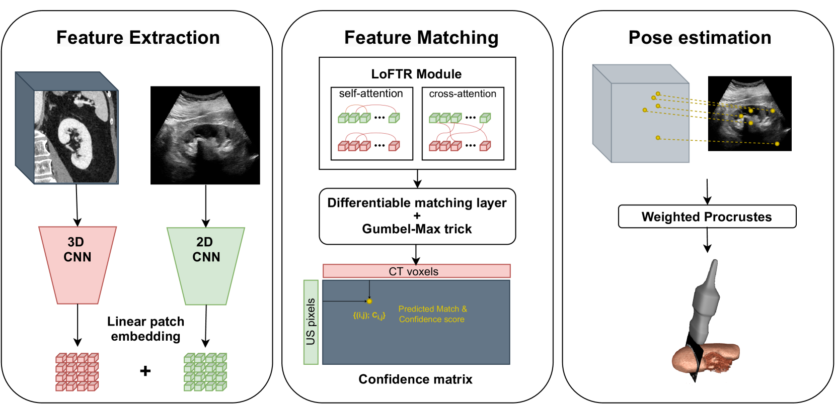
Transformer-Based Local Feature Matching for Multimodal Image Registration
Remi Delaunay, Ruisi Zhang, Filipe C. Pedrosa, Navid Feizi, Dianne Sacco, Rajni Patel, Jayender Jagadeesan
Ultrasound imaging is a cost-effective and radiation-free modality for visualizing anatomical structures in real-time, making it ideal for guiding surgical interventions. However, its limited field-of-view, speckle noise, and imaging artifacts make it difficult to interpret the images for inexperienced users. In this paper, we propose a new 2D ultrasound to 3D CT registration method to improve surgical guidance during ultrasound-guided interventions. Our approach adopts a dense feature matching method called LoFTR to our multimodal registration problem. We learn to predict dense coarse-to-fine correspondences using a Transformer-based architecture to estimate a robust rigid transformation between a 2D ultrasound frame and a CT scan. Additionally, a fully differentiable pose estimation method is introduced, optimizing LoFTR on pose estimation error during training. Experiments conducted on a multimodal dataset of ex vivo porcine kidneys demonstrate the method's promising results for intraoperative, trackerless ultrasound pose estimation. By mapping 2D ultrasound frames into the 3D CT volume space, the method provides intraoperative guidance, potentially improving surgical workflows and image interpretation.
Read more4/26/2024

0
Effective stripe artefact removal by a variational method: application to light-sheet microscopy, FIB-SEM and remote sensing images
Niklas Rottmayer, Claudia Redenbach, Florian Fahrbach
Light-sheet fluorescence microscopy (LSFM) is used to capture volume images of biological specimens. It offers high contrast deep inside densely fluorescence labelled samples, fast acquisition speed and minimal harmful effects on the sample. However, LSFM images often show strong stripe artifacts originating from light-matter interactions. We propose a robust variational method suitable for removing stripes which outperforms existing methods and offers flexibility through two adjustable parameters. This tool is widely applicable to improve visual quality as well as facilitate downstream processing and analysis of images acquired on systems that do not provide hardware-based destriping methods. An evaluation of methods is performed on LSFM, focused ion beam scanning electron microscopy (FIB-SEM) and remote sensing data, supplemented by synthetic LSFM images. The latter is obtained by simulating the imaging process on virtual samples.
Read more4/5/2024