Reliable Multi-View Learning with Conformal Prediction for Aortic Stenosis Classification in Echocardiography

0

Sign in to get full access
Overview
- Reliable multi-view learning approach for aortic stenosis classification in echocardiography
- Leverages conformal prediction to provide uncertainty estimates alongside model predictions
- Aims to improve reliability and clinical applicability of AI-based aortic stenosis diagnosis
Plain English Explanation
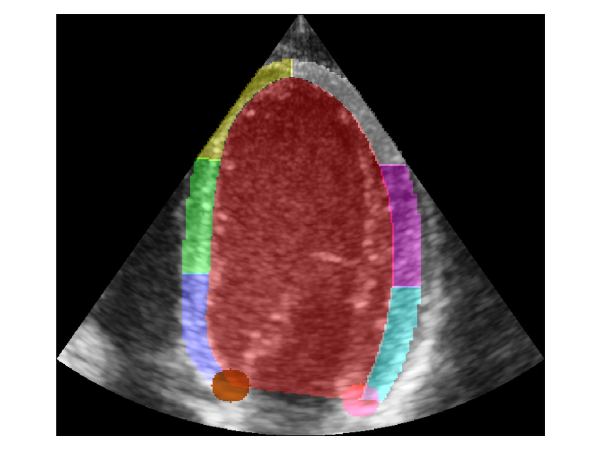
This research focuses on developing a reliable and clinically-applicable AI system for diagnosing a heart condition called aortic stenosis using echocardiography (ultrasound imaging of the heart). Aortic stenosis is a narrowing of the aortic valve that can lead to serious health problems if left untreated.
The key innovation of this work is the use of a multi-view learning approach, which combines information from multiple camera angles (views) of the heart to make a more accurate diagnosis. Additionally, the system employs conformal prediction to provide uncertainty estimates alongside the model's predictions.
This helps make the system more reliable by indicating when it is less confident in its diagnosis, which is crucial for clinical applications where incorrect diagnoses can have serious consequences.
Technical Explanation
The researchers developed a multi-view neural network architecture that takes echocardiography videos from multiple camera angles as input. This allows the model to leverage complementary information from different perspectives of the heart to improve classification accuracy.
To quantify the model's uncertainty, they used a conformal prediction framework. This involves training a separate model to estimate prediction intervals, which indicate the range of values the true label is likely to fall within. By providing these uncertainty estimates alongside the model's predictions, clinicians can better interpret the system's output and make more informed decisions.
The model was trained and evaluated on a dataset of echocardiography videos from patients with and without aortic stenosis. The results showed that the multi-view approach outperformed single-view models, and the conformal prediction intervals were well-calibrated, meaning the true labels fell within the predicted intervals at the desired confidence level.
Critical Analysis
The researchers acknowledge several limitations of their work. First, the dataset was relatively small, which may limit the model's generalization to diverse patient populations. Additionally, the conformal prediction intervals were computed independently for each view, rather than jointly considering the multi-view information.
Another potential concern is the interpretability of the multi-view neural network architecture. While the uncertainty estimates provided by conformal prediction can help, the underlying decision-making process of the model may still be opaque to clinicians. Further research is needed to explore more interpretable AI approaches for echocardiography analysis.
Overall, this work represents an important step towards reliable and clinically-useful AI systems for aortic stenosis diagnosis. The combination of multi-view learning and conformal prediction is a promising direction, but continued research and validation in larger, more diverse patient populations will be crucial for transitioning such systems into real-world clinical practice.
Conclusion
This research presents a novel approach for aortic stenosis classification in echocardiography that leverages multi-view learning and conformal prediction to improve reliability and clinical applicability. By providing uncertainty estimates alongside model predictions, the system can better support clinicians in making accurate and informed diagnoses, potentially leading to more timely and effective treatment for patients with this serious heart condition.
The work highlights the potential of advanced AI techniques to enhance medical imaging analysis, but also underscores the importance of continued research and validation to ensure the safety and trustworthiness of such systems in real-world clinical settings.
This summary was produced with help from an AI and may contain inaccuracies - check out the links to read the original source documents!
Related Papers
0
Reliable Multi-View Learning with Conformal Prediction for Aortic Stenosis Classification in Echocardiography
Ang Nan Gu, Michael Tsang, Hooman Vaseli, Teresa Tsang, Purang Abolmaesumi
The fundamental problem with ultrasound-guided diagnosis is that the acquired images are often 2-D cross-sections of a 3-D anatomy, potentially missing important anatomical details. This limitation leads to challenges in ultrasound echocardiography, such as poor visualization of heart valves or foreshortening of ventricles. Clinicians must interpret these images with inherent uncertainty, a nuance absent in machine learning's one-hot labels. We propose Re-Training for Uncertainty (RT4U), a data-centric method to introduce uncertainty to weakly informative inputs in the training set. This simple approach can be incorporated to existing state-of-the-art aortic stenosis classification methods to further improve their accuracy. When combined with conformal prediction techniques, RT4U can yield adaptively sized prediction sets which are guaranteed to contain the ground truth class to a high accuracy. We validate the effectiveness of RT4U on three diverse datasets: a public (TMED-2) and a private AS dataset, along with a CIFAR-10-derived toy dataset. Results show improvement on all the datasets.
Read more9/17/2024
👨🏫
0
Detecting Heart Disease from Multi-View Ultrasound Images via Supervised Attention Multiple Instance Learning
Zhe Huang, Benjamin S. Wessler, Michael C. Hughes
Aortic stenosis (AS) is a degenerative valve condition that causes substantial morbidity and mortality. This condition is under-diagnosed and under-treated. In clinical practice, AS is diagnosed with expert review of transthoracic echocardiography, which produces dozens of ultrasound images of the heart. Only some of these views show the aortic valve. To automate screening for AS, deep networks must learn to mimic a human expert's ability to identify views of the aortic valve then aggregate across these relevant images to produce a study-level diagnosis. We find previous approaches to AS detection yield insufficient accuracy due to relying on inflexible averages across images. We further find that off-the-shelf attention-based multiple instance learning (MIL) performs poorly. We contribute a new end-to-end MIL approach with two key methodological innovations. First, a supervised attention technique guides the learned attention mechanism to favor relevant views. Second, a novel self-supervised pretraining strategy applies contrastive learning on the representation of the whole study instead of individual images as commonly done in prior literature. Experiments on an open-access dataset and an external validation set show that our approach yields higher accuracy while reducing model size.
Read more4/8/2024

0
Multimodal Fusion of Echocardiography and Electronic Health Records for the Detection of Cardiac Amyloidosis
Zishun Feng, Joseph A. Sivak, Ashok K. Krishnamurthy
Cardiac amyloidosis, a rare and highly morbid condition, presents significant challenges for detection through echocardiography. Recently, there has been a surge in proposing machine-learning algorithms to identify cardiac amyloidosis, with the majority being imaging-based deep-learning approaches that require extensive data. In this study, we introduce a novel transformer-based multimodal fusion algorithm that leverages information from both imaging and electronic health records. Specifically, our approach utilizes echocardiography videos from both the parasternal long-axis (PLAX) view and the apical 4-chamber (A4C) view along with patients' demographic data, laboratory tests, and cardiac metrics to predict the probability of cardiac amyloidosis. We evaluated our method using 5-fold cross-validation on a dataset comprising 41 patients and achieved an Area Under the Receiver Operating Characteristic curve (AUROC) of 0.94. The experimental results demonstrate that our approach can achieve competitive results with a significantly smaller dataset compared to prior imaging-based methods that required data from thousands of patients. This underscores the potential of leveraging multimodal data to enhance diagnostic accuracy in the identification of complex cardiac conditions such as cardiac amyloidosis.
Read more6/10/2024
0
Regional quality estimation for echocardiography using deep learning
Gilles Van De Vyver, Svein-Erik M{aa}s{o}y, H{aa}vard Dalen, Bj{o}rnar Leangen Grenne, Espen Holte, Sindre Hellum Olaisen, John Nyberg, Andreas {O}stvik, Lasse L{o}vstakken, Erik Smistad
Automatic estimation of cardiac ultrasound image quality can be beneficial for guiding operators and ensuring the accuracy of clinical measurements. Previous work often fails to distinguish the view correctness of the echocardiogram from the image quality. Additionally, previous studies only provide a global image quality value, which limits their practical utility. In this work, we developed and compared three methods to estimate image quality: 1) classic pixel-based metrics like the generalized contrast-to-noise ratio (gCNR) on myocardial segments as region of interest and left ventricle lumen as background, obtained using a U-Net segmentation 2) local image coherence derived from a U-Net model that predicts coherence from B-Mode images 3) a deep convolutional network that predicts the quality of each region directly in an end-to-end fashion. We evaluate each method against manual regional image quality annotations by three experienced cardiologists. The results indicate poor performance of the gCNR metric, with Spearman correlation to the annotations of rho = 0.24. The end-to-end learning model obtains the best result, rho = 0.69, comparable to the inter-observer correlation, rho = 0.63. Finally, the coherence-based method, with rho = 0.58, outperformed the classical metrics and is more generic than the end-to-end approach.
Read more8/28/2024