Revisiting Adaptive Cellular Recognition Under Domain Shifts: A Contextual Correspondence View

0
Sign in to get full access
Overview
- The paper explores adaptive cellular recognition under domain shifts, using a contextual correspondence view.
- It examines how machine learning models can effectively recognize cellular structures across different imaging conditions or "domains".
- The research proposes a novel approach that leverages contextual information and correspondence between domains to improve the model's performance.
- The method is evaluated on various cellular recognition tasks, demonstrating its effectiveness in handling domain shifts.
Plain English Explanation
Recognizing and classifying different types of cells in microscopic images is an important task in biology and medicine. However, this can be challenging when the images come from different sources or "domains" - for example, cells imaged under different lighting conditions, using different microscopes, or in different laboratories.
The researchers in this paper developed a new way to help AI models adapt to these changes in the imaging "domain". Their approach focuses on the context around the cells, not just the cells themselves. By understanding how the cells relate to their surrounding environment, the model can better recognize the same cell types even when the overall image looks quite different.
The key insight is that there are often consistent relationships or "correspondences" between the features in different imaging domains. For example, even if the color and brightness of an image changes, the spatial arrangement of the cells and other structures may remain similar. By capturing these contextual correspondences, the model can learn to "translate" between the different imaging domains and recognize the same cellular structures.
The researchers tested their method on several cell recognition tasks and found that it outperformed previous approaches, especially when the training and test data came from quite different domains. This is an important step forward in making AI-powered cellular analysis more robust and reliable across diverse imaging conditions.
Technical Explanation
The paper proposes a "Contextual Correspondence Learning" (CCL) framework to address the problem of adaptive cellular recognition under domain shifts. The key components of CCL are:
-
Contextual Feature Extraction: The model extracts features not just from the target cells, but also from the surrounding contextual regions. This allows the model to capture spatial relationships and other contextual cues that are often preserved across imaging domains.
-
Cross-Domain Correspondence Learning: The model learns to establish correspondence between the contextual features extracted from different imaging domains. This enables the model to "translate" between the feature representations of the source and target domains.
-
Adaptive Classification: The model uses the learned cross-domain correspondences to adapt its classification head, allowing it to accurately recognize cell types even when the input comes from a different imaging domain than the training data.
The paper evaluates CCL on several cellular recognition tasks, including nucleus segmentation, cell type classification, and tissue segmentation. The experiments demonstrate that CCL outperforms previous state-of-the-art domain adaptation methods, particularly when there is a significant shift between the training and test domains.
Critical Analysis
The paper presents a well-designed and thorough study, with a clear motivation and a novel technical approach. The key strength of the work is the emphasis on capturing contextual information and establishing cross-domain correspondences, which appears to be an effective strategy for handling domain shifts in cellular recognition tasks.
However, the paper does not address some potential limitations of the proposed approach. For example, the method may struggle with significant changes in the underlying cellular morphology or structure across domains, beyond just the imaging conditions. Additionally, the computational complexity of the correspondence learning process could be a concern for real-world deployment, especially for large-scale cellular datasets.
Further research could explore ways to make the correspondence learning more efficient, as well as investigate the robustness of the method to more extreme domain shifts or changes in the fundamental cellular properties. Incorporating unsupervised latent stain adaptation or weakly supervised object detection techniques could also be a promising direction to further improve the model's adaptability.
Conclusion
This paper presents a novel approach, Contextual Correspondence Learning (CCL), for addressing the challenge of adaptive cellular recognition under domain shifts. By focusing on the contextual information around the target cells and learning cross-domain correspondences, the method demonstrates significant improvements over previous state-of-the-art techniques.
The work has important implications for advancing the reliability and robustness of AI-powered cellular analysis, which is crucial for applications in biology, medicine, and beyond. As the field of computational pathology continues to evolve, techniques like CCL that can handle domain shifts will become increasingly valuable for translating these powerful tools into real-world clinical and research settings.
This summary was produced with help from an AI and may contain inaccuracies - check out the links to read the original source documents!
Related Papers
0
Revisiting Adaptive Cellular Recognition Under Domain Shifts: A Contextual Correspondence View
Jianan Fan, Dongnan Liu, Canran Li, Hang Chang, Heng Huang, Filip Braet, Mei Chen, Weidong Cai
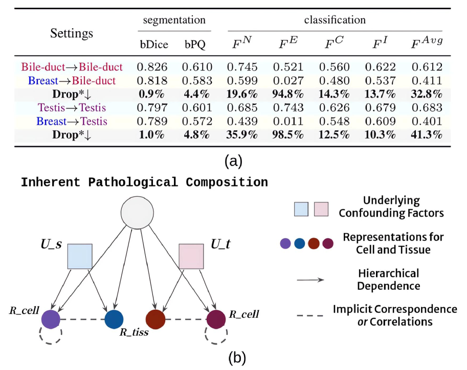
Cellular nuclei recognition serves as a fundamental and essential step in the workflow of digital pathology. However, with disparate source organs and staining procedures among histology image clusters, the scanned tiles inherently conform to a non-uniform data distribution, which induces deteriorated promises for general cross-cohort usages. Despite the latest efforts leveraging domain adaptation to mitigate distributional discrepancy, those methods are subjected to modeling the morphological characteristics of each cell individually, disregarding the hierarchical latent structure and intrinsic contextual correspondences across the tumor micro-environment. In this work, we identify the importance of implicit correspondences across biological contexts for exploiting domain-invariant pathological composition and thereby propose to exploit the dependence over various biological structures for domain adaptive cellular recognition. We discover those high-level correspondences via unsupervised contextual modeling and use them as bridges to facilitate adaptation over diverse organs and stains. In addition, to further exploit the rich spatial contexts embedded amongst nuclear communities, we propose self-adaptive dynamic distillation to secure instance-aware trade-offs across different model constituents. The proposed method is extensively evaluated on a broad spectrum of cross-domain settings under miscellaneous data distribution shifts and outperforms the state-of-the-art methods by a substantial margin. Code is available at https://github.com/camwew/CellularRecognition_DA_CC.
Read more7/22/2024

0
Mix-Domain Contrastive Learning for Unpaired H&E-to-IHC Stain Translation
Song Wang, Zhong Zhang, Huan Yan, Ming Xu, Guanghui Wang
H&E-to-IHC stain translation techniques offer a promising solution for precise cancer diagnosis, especially in low-resource regions where there is a shortage of health professionals and limited access to expensive equipment. Considering the pixel-level misalignment of H&E-IHC image pairs, current research explores the pathological consistency between patches from the same positions of the image pair. However, most of them overemphasize the correspondence between domains or patches, overlooking the side information provided by the non-corresponding objects. In this paper, we propose a Mix-Domain Contrastive Learning (MDCL) method to leverage the supervision information in unpaired H&E-to-IHC stain translation. Specifically, the proposed MDCL method aggregates the inter-domain and intra-domain pathology information by estimating the correlation between the anchor patch and all the patches from the matching images, encouraging the network to learn additional contrastive knowledge from mixed domains. With the mix-domain pathology information aggregation, MDCL enhances the pathological consistency between the corresponding patches and the component discrepancy of the patches from the different positions of the generated IHC image. Extensive experiments on two H&E-to-IHC stain translation datasets, namely MIST and BCI, demonstrate that the proposed method achieves state-of-the-art performance across multiple metrics.
Read more9/4/2024

0
Co-synthesis of Histopathology Nuclei Image-Label Pairs using a Context-Conditioned Joint Diffusion Model
Seonghui Min, Hyun-Jic Oh, Won-Ki Jeong
In multi-class histopathology nuclei analysis tasks, the lack of training data becomes a main bottleneck for the performance of learning-based methods. To tackle this challenge, previous methods have utilized generative models to increase data by generating synthetic samples. However, existing methods often overlook the importance of considering the context of biological tissues (e.g., shape, spatial layout, and tissue type) in the synthetic data. Moreover, while generative models have shown superior performance in synthesizing realistic histopathology images, none of the existing methods are capable of producing image-label pairs at the same time. In this paper, we introduce a novel framework for co-synthesizing histopathology nuclei images and paired semantic labels using a context-conditioned joint diffusion model. We propose conditioning of a diffusion model using nucleus centroid layouts with structure-related text prompts to incorporate spatial and structural context information into the generation targets. Moreover, we enhance the granularity of our synthesized semantic labels by generating instance-wise nuclei labels using distance maps synthesized concurrently in conjunction with the images and semantic labels. We demonstrate the effectiveness of our framework in generating high-quality samples on multi-institutional, multi-organ, and multi-modality datasets. Our synthetic data consistently outperforms existing augmentation methods in the downstream tasks of nuclei segmentation and classification.
Read more9/5/2024

0
Source-Free Domain Adaptation of Weakly-Supervised Object Localization Models for Histology
Alexis Guichemerre, Soufiane Belharbi, Tsiry Mayet, Shakeeb Murtaza, Pourya Shamsolmoali, Luke McCaffrey, Eric Granger
Given the emergence of deep learning, digital pathology has gained popularity for cancer diagnosis based on histology images. Deep weakly supervised object localization (WSOL) models can be trained to classify histology images according to cancer grade and identify regions of interest (ROIs) for interpretation, using inexpensive global image-class annotations. A WSOL model initially trained on some labeled source image data can be adapted using unlabeled target data in cases of significant domain shifts caused by variations in staining, scanners, and cancer type. In this paper, we focus on source-free (unsupervised) domain adaptation (SFDA), a challenging problem where a pre-trained source model is adapted to a new target domain without using any source domain data for privacy and efficiency reasons. SFDA of WSOL models raises several challenges in histology, most notably because they are not intended to adapt for both classification and localization tasks. In this paper, 4 state-of-the-art SFDA methods, each one representative of a main SFDA family, are compared for WSOL in terms of classification and localization accuracy. They are the SFDA-Distribution Estimation, Source HypOthesis Transfer, Cross-Domain Contrastive Learning, and Adaptively Domain Statistics Alignment. Experimental results on the challenging Glas (smaller, breast cancer) and Camelyon16 (larger, colon cancer) histology datasets indicate that these SFDA methods typically perform poorly for localization after adaptation when optimized for classification.
Read more5/14/2024