Segmentation of Prostate Tumour Volumes from PET Images is a Different Ball Game

0
✨
Sign in to get full access
Overview
- This technical appendix provides details on the dataset and code used in the research paper "Segmentation of Prostate Tumour Volumes from PET Images is a Different Ball Game".
- The research focused on developing deep learning-based algorithms for accurately segmenting prostate tumors from PET images, which is a challenging task.
- The authors collected a unique dataset of PET scans and manual tumor segmentations, and developed custom neural network architectures to tackle this problem.
Plain English Explanation
The research paper you provided describes a study that aimed to improve the way doctors can identify and measure prostate tumors using PET imaging scans. PET scans use a radioactive tracer to highlight areas of the body with higher metabolic activity, which can indicate the presence of tumors.
However, accurately segmenting, or outlining, prostate tumors in PET images is quite difficult, as the tumors can be small and the images can be noisy. The researchers in this study developed specialized deep learning models - a type of artificial intelligence - to try to automate this tumor segmentation process and make it more accurate and reliable.
They started by collecting a unique dataset of PET scans along with "ground truth" manual segmentations of the tumors, provided by expert radiologists. This allowed them to train and test their deep learning models on real patient data. [link to https://aimodels.fyi/papers/arxiv/deep-learning-based-segmentation-tumors-petct-volumes]
The researchers then experimented with different neural network architectures and training approaches to see which ones could most accurately locate and outline the prostate tumors in the PET images. This involved a lot of trial and error to find the right balance of model complexity, training data, and optimization techniques. [link to https://aimodels.fyi/papers/arxiv/segmentation-free-outcome-prediction-head-neck-cancer]
Ultimately, the researchers were able to develop deep learning models that could segment prostate tumors from PET scans with a high degree of accuracy, outperforming traditional segmentation methods. This has important implications for improving prostate cancer diagnosis and monitoring, as it could help doctors more precisely measure and track tumors over time. [link to https://aimodels.fyi/papers/arxiv/deep-learning-segmentation-small-volumes-ct-images]
Technical Explanation
The key elements of this research paper are:
-
Dataset Collection: The authors compiled a unique dataset of 68 PET/CT scans of prostate cancer patients, along with manual tumor segmentations provided by expert radiologists. This "ground truth" data was crucial for training and evaluating the deep learning models. [link to https://aimodels.fyi/papers/arxiv/ai-based-automatic-segmentation-prostate-multi-modality]
-
Model Architecture: The researchers experimented with several customized convolutional neural network (CNN) architectures, including a U-Net-based model and a Siamese network design. These models were trained to take in the 3D PET/CT volumes and output segmented prostate tumor regions. [link to https://aimodels.fyi/papers/arxiv/towards-ai-lesion-tracking-petct-imaging-siamese]
-
Training and Optimization: The authors explored various training techniques, such as data augmentation, transfer learning, and multi-task learning, to improve the models' performance on the challenging prostate tumor segmentation task. They also utilized loss functions and metrics tailored for the medical imaging domain.
-
Evaluation: The trained models were evaluated on held-out test data using standard segmentation metrics, such as Dice coefficient and Hausdorff distance. The deep learning approaches were shown to outperform traditional segmentation methods, particularly for small and irregularly shaped tumors.
Critical Analysis
The authors acknowledge several limitations and areas for future research in this work:
-
The dataset, while unique, is still relatively small, which may limit the generalizability of the models. Expanding the dataset with more patient data could help improve the models' performance and robustness. [link to https://aimodels.fyi/papers/arxiv/deep-learning-based-segmentation-tumors-petct-volumes]
-
The manual tumor segmentations provided by radiologists may contain some inherent variability and uncertainty, which could introduce noise into the training data. Exploring ways to account for this annotation uncertainty could be beneficial. [link to https://aimodels.fyi/papers/arxiv/segmentation-free-outcome-prediction-head-neck-cancer]
-
The models were trained and evaluated on PET/CT scans, but the authors note that PET imaging alone may be sufficient for tumor segmentation in many cases. Investigating the use of PET-only models could simplify the clinical workflow and reduce patient exposure to radiation. [link to https://aimodels.fyi/papers/arxiv/deep-learning-segmentation-small-volumes-ct-images]
-
The clinical impact and practical implementation of these deep learning-based segmentation tools in real-world prostate cancer care settings were not directly addressed in this paper. Further research is needed to demonstrate the tangible benefits to patients and healthcare providers. [link to https://aimodels.fyi/papers/arxiv/ai-based-automatic-segmentation-prostate-multi-modality, https://aimodels.fyi/papers/arxiv/towards-ai-lesion-tracking-petct-imaging-siamese]
Conclusion
This technical appendix provides valuable details on the dataset and custom deep learning models developed to address the challenging task of prostate tumor segmentation from PET images. The authors have made significant progress in this area, demonstrating the potential of AI-powered medical image analysis to improve prostate cancer diagnosis and monitoring. However, further research is needed to address the limitations and translate these advancements into real-world clinical practice. The insights gained from this work could also inform the development of similar deep learning techniques for segmenting tumors in other anatomical regions and medical imaging modalities.
This summary was produced with help from an AI and may contain inaccuracies - check out the links to read the original source documents!
Related Papers
✨
0
Segmentation of Prostate Tumour Volumes from PET Images is a Different Ball Game
Shrajan Bhandary, Dejan Kuhn, Zahra Babaiee, Tobias Fechter, Simon K. B. Spohn, Constantinos Zamboglou, Anca-Ligia Grosu, Radu Grosu
Accurate segmentation of prostate tumours from PET images presents a formidable challenge in medical image analysis. Despite considerable work and improvement in delineating organs from CT and MR modalities, the existing standards do not transfer well and produce quality results in PET related tasks. Particularly, contemporary methods fail to accurately consider the intensity-based scaling applied by the physicians during manual annotation of tumour contours. In this paper, we observe that the prostate-localised uptake threshold ranges are beneficial for suppressing outliers. Therefore, we utilize the intensity threshold values, to implement a new custom-feature-clipping normalisation technique. We evaluate multiple, established U-Net variants under different normalisation schemes, using the nnU-Net framework. All models were trained and tested on multiple datasets, obtained with two radioactive tracers: [68-Ga]Ga-PSMA-11 and [18-F]PSMA-1007. Our results show that the U-Net models achieve much better performance when the PET scans are preprocessed with our novel clipping technique.
Read more7/16/2024
0
How To Segment in 3D Using 2D Models: Automated 3D Segmentation of Prostate Cancer Metastatic Lesions on PET Volumes Using Multi-Angle Maximum Intensity Projections and Diffusion Models
Amirhosein Toosi, Sara Harsini, Franc{c}ois B'enard, Carlos Uribe, Arman Rahmim
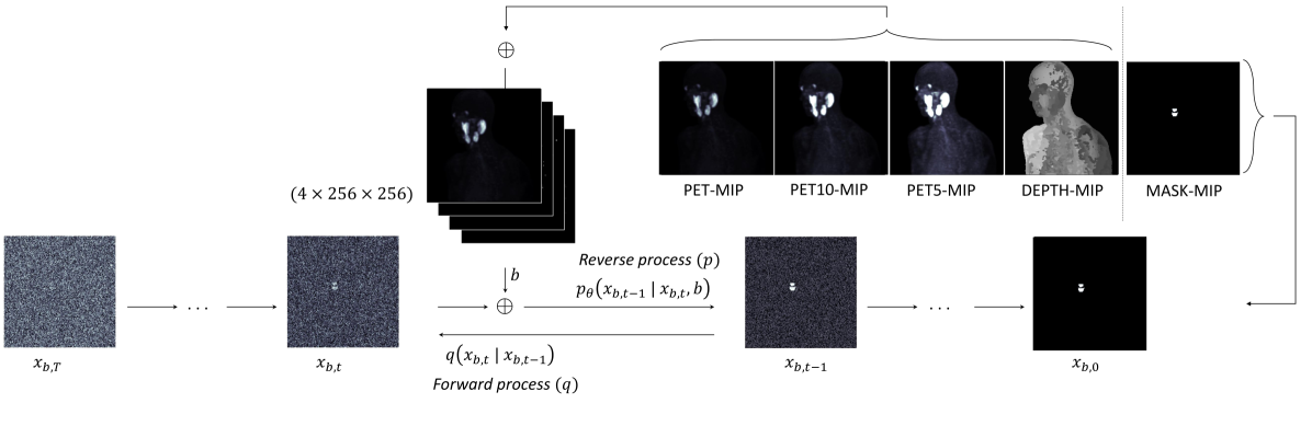
Prostate specific membrane antigen (PSMA) positron emission tomography/computed tomography (PET/CT) imaging provides a tremendously exciting frontier in visualization of prostate cancer (PCa) metastatic lesions. However, accurate segmentation of metastatic lesions is challenging due to low signal-to-noise ratios and variable sizes, shapes, and locations of the lesions. This study proposes a novel approach for automated segmentation of metastatic lesions in PSMA PET/CT 3D volumetric images using 2D denoising diffusion probabilistic models (DDPMs). Instead of 2D trans-axial slices or 3D volumes, the proposed approach segments the lesions on generated multi-angle maximum intensity projections (MA-MIPs) of the PSMA PET images, then obtains the final 3D segmentation masks from 3D ordered subset expectation maximization (OSEM) reconstruction of 2D MA-MIPs segmentations. Our proposed method achieved superior performance compared to state-of-the-art 3D segmentation approaches in terms of accuracy and robustness in detecting and segmenting small metastatic PCa lesions. The proposed method has significant potential as a tool for quantitative analysis of metastatic burden in PCa patients.
Read more7/29/2024

0
Deep Learning-Based Segmentation of Tumors in PET/CT Volumes: Benchmark of Different Architectures and Training Strategies
Monika G'orka, Daniel Jaworek, Marek Wodzinski
Cancer is one of the leading causes of death globally, and early diagnosis is crucial for patient survival. Deep learning algorithms have great potential for automatic cancer analysis. Artificial intelligence has achieved high performance in recognizing and segmenting single lesions. However, diagnosing multiple lesions remains a challenge. This study examines and compares various neural network architectures and training strategies for automatically segmentation of cancer lesions using PET/CT images from the head, neck, and whole body. The authors analyzed datasets from the AutoPET and HECKTOR challenges, exploring popular single-step segmentation architectures and presenting a two-step approach. The results indicate that the V-Net and nnU-Net models were the most effective for their respective datasets. The results for the HECKTOR dataset ranged from 0.75 to 0.76 for the aggregated Dice coefficient. Eliminating cancer-free cases from the AutoPET dataset was found to improve the performance of most models. In the case of AutoPET data, the average segmentation efficiency after training only on images containing cancer lesions increased from 0.55 to 0.66 for the classic Dice coefficient and from 0.65 to 0.73 for the aggregated Dice coefficient. The research demonstrates the potential of artificial intelligence in precise oncological diagnostics and may contribute to the development of more targeted and effective cancer assessment techniques.
Read more4/16/2024

0
New!Enhancing Lesion Segmentation in PET/CT Imaging with Deep Learning and Advanced Data Preprocessing Techniques
Jiayi Liu, Qiaoyi Xue, Youdan Feng, Tianming Xu, Kaixin Shen, Chuyun Shen, Yuhang Shi
The escalating global cancer burden underscores the critical need for precise diagnostic tools in oncology. This research employs deep learning to enhance lesion segmentation in PET/CT imaging, utilizing a dataset of 900 whole-body FDG-PET/CT and 600 PSMA-PET/CT studies from the AutoPET challenge III. Our methodical approach includes robust preprocessing and data augmentation techniques to ensure model robustness and generalizability. We investigate the influence of non-zero normalization and modifications to the data augmentation pipeline, such as the introduction of RandGaussianSharpen and adjustments to the Gamma transform parameter. This study aims to contribute to the standardization of preprocessing and augmentation strategies in PET/CT imaging, potentially improving the diagnostic accuracy and the personalized management of cancer patients. Our code will be open-sourced and available at https://github.com/jiayiliu-pku/DC2024.
Read more9/17/2024