Segmentation Quality and Volumetric Accuracy in Medical Imaging

0
Sign in to get full access
Overview
- This paper investigates the quality and accuracy of medical image segmentation techniques, which are critical for various clinical applications.
- It explores how different evaluation metrics and uncertainty estimation methods can impact the assessment of segmentation performance.
- The research presents a comprehensive study of these aspects, aiming to provide insights for improving the real-world applicability of medical image segmentation algorithms.
Plain English Explanation
Medical imaging plays a crucial role in healthcare, allowing doctors to diagnose and monitor various conditions. One important aspect of medical imaging is image segmentation, which involves dividing the image into distinct regions or structures, such as organs, tumors, or blood vessels. Accurate segmentation is essential for many clinical applications, such as surgical planning, disease monitoring, and treatment evaluation.
This paper examines the quality and accuracy of medical image segmentation techniques. It investigates how different evaluation metrics and uncertainty estimation methods can affect the assessment of segmentation performance. Evaluation metrics are used to quantify the accuracy of segmentation results, while uncertainty estimation methods aim to provide a measure of the reliability or confidence in the segmentation.
The research presents a comprehensive study of these aspects, with the goal of improving the real-world applicability of medical image segmentation algorithms. By understanding the impact of different evaluation approaches and uncertainty estimation techniques, the authors aim to provide insights that can help developers and clinicians better assess and trust the results of medical image segmentation algorithms in practical settings.
Technical Explanation
The paper explores the relationship between segmentation quality, volumetric accuracy, and uncertainty estimation in medical imaging. It investigates how the choice of evaluation metrics and uncertainty estimation methods can impact the assessment of segmentation performance.
The authors conduct a series of experiments using various segmentation algorithms and medical imaging datasets. They compare different evaluation metrics, such as Dice similarity coefficient and Hausdorff distance, to assess the segmentation quality. Additionally, they explore the use of uncertainty-aware evidential fusion and other uncertainty estimation techniques to quantify the reliability of the segmentation results.
The findings of the study highlight the importance of considering both segmentation quality and volumetric accuracy when evaluating medical image segmentation algorithms. The authors also emphasize the need to carefully select appropriate evaluation metrics and uncertainty estimation methods, as these choices can significantly impact the assessment of segmentation performance and the real-world applicability of the algorithms.
Critical Analysis
The paper provides a comprehensive and well-designed study, addressing an important topic in the field of medical image segmentation. The authors have carefully considered various evaluation metrics and uncertainty estimation techniques, and their findings offer valuable insights for the research community and practitioners.
However, the paper also acknowledges certain limitations and areas for further research. For example, the authors mention that the study was conducted on a limited set of medical imaging datasets and segmentation algorithms. Expanding the scope to a broader range of datasets and algorithms could further strengthen the generalizability of the findings.
Additionally, the paper does not delve into the potential sources of uncertainty in medical image segmentation, such as image acquisition artifacts, anatomical variability, or inconsistencies in ground truth labeling. Exploring these factors and their impact on segmentation performance could provide a more holistic understanding of the challenges in this domain.
Furthermore, the paper could benefit from a more in-depth discussion of the practical implications of the findings, particularly for clinical decision-making and the development of reliable medical image analysis tools. Addressing these aspects could enhance the relevance and impact of the research.
Conclusion
This paper offers a valuable contribution to the field of medical image segmentation by investigating the relationship between segmentation quality, volumetric accuracy, and uncertainty estimation. The authors' comprehensive study highlights the importance of carefully selecting evaluation metrics and uncertainty estimation methods to better assess the real-world applicability of segmentation algorithms.
The findings from this research can inform the development of more robust and reliable medical image analysis tools, ultimately supporting clinicians in making informed decisions and improving patient outcomes. The insights provided in this paper serve as a valuable resource for the research community and practitioners working in the field of medical imaging and computer-assisted diagnosis.
This summary was produced with help from an AI and may contain inaccuracies - check out the links to read the original source documents!
Related Papers
0
Segmentation Quality and Volumetric Accuracy in Medical Imaging
Zheyuan Zhang, Ulas Bagci
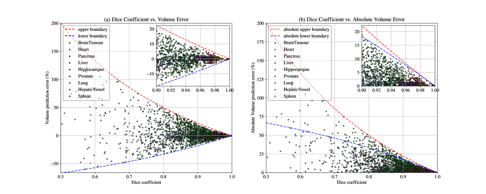
Current medical image segmentation relies on the region-based (Dice, F1-score) and boundary-based (Hausdorff distance, surface distance) metrics as the de-facto standard. While these metrics are widely used, they lack a unified interpretation, particularly regarding volume agreement. Clinicians often lack clear benchmarks to gauge the goodness of segmentation results based on these metrics. Recognizing the clinical relevance of volumetry, we utilize relative volume prediction error (vpe) to directly assess the accuracy of volume predictions derived from segmentation tasks. Our work integrates theoretical analysis and empirical validation across diverse datasets. We delve into the often-ambiguous relationship between segmentation quality (measured by Dice) and volumetric accuracy in clinical practice. Our findings highlight the critical role of incorporating volumetric prediction accuracy into segmentation evaluation. This approach empowers clinicians with a more nuanced understanding of segmentation performance, ultimately improving the interpretation and utility of these metrics in real-world healthcare settings.
Read more5/15/2024
🖼️
0
SegVol: Universal and Interactive Volumetric Medical Image Segmentation
Yuxin Du, Fan Bai, Tiejun Huang, Bo Zhao
Precise image segmentation provides clinical study with instructive information. Despite the remarkable progress achieved in medical image segmentation, there is still an absence of a 3D foundation segmentation model that can segment a wide range of anatomical categories with easy user interaction. In this paper, we propose a 3D foundation segmentation model, named SegVol, supporting universal and interactive volumetric medical image segmentation. By scaling up training data to 90K unlabeled Computed Tomography (CT) volumes and 6K labeled CT volumes, this foundation model supports the segmentation of over 200 anatomical categories using semantic and spatial prompts. To facilitate efficient and precise inference on volumetric images, we design a zoom-out-zoom-in mechanism. Extensive experiments on 22 anatomical segmentation tasks verify that SegVol outperforms the competitors in 19 tasks, with improvements up to 37.24% compared to the runner-up methods. We demonstrate the effectiveness and importance of specific designs by ablation study. We expect this foundation model can promote the development of volumetric medical image analysis. The model and code are publicly available at: https://github.com/BAAI-DCAI/SegVol.
Read more8/30/2024

0
Comparative Benchmarking of Failure Detection Methods in Medical Image Segmentation: Unveiling the Role of Confidence Aggregation
Maximilian Zenk, David Zimmerer, Fabian Isensee, Jeremias Traub, Tobias Norajitra, Paul F. Jager, Klaus Maier-Hein
Semantic segmentation is an essential component of medical image analysis research, with recent deep learning algorithms offering out-of-the-box applicability across diverse datasets. Despite these advancements, segmentation failures remain a significant concern for real-world clinical applications, necessitating reliable detection mechanisms. This paper introduces a comprehensive benchmarking framework aimed at evaluating failure detection methodologies within medical image segmentation. Through our analysis, we identify the strengths and limitations of current failure detection metrics, advocating for the risk-coverage analysis as a holistic evaluation approach. Utilizing a collective dataset comprising five public 3D medical image collections, we assess the efficacy of various failure detection strategies under realistic test-time distribution shifts. Our findings highlight the importance of pixel confidence aggregation and we observe superior performance of the pairwise Dice score (Roy et al., 2019) between ensemble predictions, positioning it as a simple and robust baseline for failure detection in medical image segmentation. To promote ongoing research, we make the benchmarking framework available to the community.
Read more6/6/2024

0
Robust Conformal Volume Estimation in 3D Medical Images
Benjamin Lambert, Florence Forbes, Senan Doyle, Michel Dojat
Volumetry is one of the principal downstream applications of 3D medical image segmentation, for example, to detect abnormal tissue growth or for surgery planning. Conformal Prediction is a promising framework for uncertainty quantification, providing calibrated predictive intervals associated with automatic volume measurements. However, this methodology is based on the hypothesis that calibration and test samples are exchangeable, an assumption that is in practice often violated in medical image applications. A weighted formulation of Conformal Prediction can be framed to mitigate this issue, but its empirical investigation in the medical domain is still lacking. A potential reason is that it relies on the estimation of the density ratio between the calibration and test distributions, which is likely to be intractable in scenarios involving high-dimensional data. To circumvent this, we propose an efficient approach for density ratio estimation relying on the compressed latent representations generated by the segmentation model. Our experiments demonstrate the efficiency of our approach to reduce the coverage error in the presence of covariate shifts, in both synthetic and real-world settings. Our implementation is available at https://github.com/benolmbrt/wcp_miccai
Read more7/30/2024