Self-supervised learning for classifying paranasal anomalies in the maxillary sinus

0

Sign in to get full access
Overview
- This paper presents a self-supervised learning approach for classifying paranasal anomalies in the maxillary sinus using medical imaging data.
- The researchers developed a model that can automatically detect and classify various types of abnormalities in the maxillary sinus, a key area for diagnosing sinus-related conditions.
- The self-supervised learning technique allows the model to learn useful features from the data without requiring extensive manual labeling, making it more scalable and efficient compared to traditional supervised approaches.
Plain English Explanation
The maxillary sinus is a hollow space in the cheekbone that can sometimes develop abnormalities or anomalies. These issues can be indicators of various sinus-related health problems, so being able to accurately identify and classify them is important for medical diagnosis and treatment.
The researchers in this study developed a new AI-powered tool that can automatically detect and categorize different types of paranasal anomalies in the maxillary sinus based on medical imaging data, such as CT scans or MRI images. Promoting internal link Unlike traditional machine learning approaches that require a lot of manually labeled training data, this tool uses a self-supervised learning technique. This means the model can learn useful features and patterns from the data on its own, without needing extensive human annotation.
Promoting internal link The advantage of this self-supervised approach is that it can be applied to a wider range of medical imaging data, making the tool more scalable and efficient compared to models that rely on manually curated datasets. By automatically detecting and classifying sinus anomalies, this technology could help healthcare providers make more accurate diagnoses and determine the appropriate treatments for patients with sinus-related conditions.
Technical Explanation
The researchers employed a self-supervised learning framework to train a deep neural network model for the task of classifying paranasal anomalies in the maxillary sinus. Promoting internal link The model was trained on a large dataset of CT scan images, with the self-supervised objective of learning to predict the relative positions of randomly sampled image patches. This allowed the model to learn meaningful image representations without the need for manually labeled data.
Promoting internal link The learned representations were then fine-tuned on a smaller dataset of manually annotated CT scans, where the model was trained to classify different types of paranasal anomalies in the maxillary sinus. The researchers evaluated the model's performance on a held-out test set and compared it to various baseline approaches, demonstrating the advantages of the self-supervised pre-training strategy.
Promoting internal link The results show that the self-supervised model achieved superior classification accuracy compared to fully supervised models, particularly when the labeled training data was limited. This highlights the potential of self-supervised learning techniques to enhance medical image analysis in scenarios where annotated data is scarce.
Critical Analysis
The paper provides a compelling demonstration of the benefits of self-supervised learning for medical image analysis tasks. The researchers have shown that their approach can effectively leverage large volumes of unlabeled data to learn useful representations, which can then be fine-tuned for specific classification objectives with improved performance compared to fully supervised models.
However, the paper does not address certain limitations and potential concerns. For example, the study only focuses on classifying paranasal anomalies in the maxillary sinus, and it is unclear how well the self-supervised model would generalize to other types of medical imaging tasks or anatomical regions. Additionally, the paper does not discuss the interpretability or explainability of the model's predictions, which is an important consideration for medical applications where clinicians need to understand the reasoning behind the model's decisions.
Promoting internal link Further research could explore the performance of this self-supervised approach on a broader range of medical imaging tasks, as well as investigate ways to improve the model's interpretability and robustness to ensure its safe and effective deployment in real-world healthcare settings.
Conclusion
This paper presents a promising self-supervised learning approach for classifying paranasal anomalies in the maxillary sinus using medical imaging data. The key advantage of this technique is its ability to learn useful features from large volumes of unlabeled data, which can then be effectively fine-tuned for specific classification tasks with limited labeled data.
Promoting internal link If further developed and validated, this self-supervised model could potentially contribute to more accurate and efficient diagnosis of sinus-related conditions, ultimately leading to better patient outcomes. The approach showcases the potential of self-supervised learning to enhance medical image analysis and address the challenges of data scarcity in healthcare applications.
This summary was produced with help from an AI and may contain inaccuracies - check out the links to read the original source documents!
Related Papers
0
Self-supervised learning for classifying paranasal anomalies in the maxillary sinus
Debayan Bhattacharya, Finn Behrendt, Benjamin Tobias Becker, Lennart Maack, Dirk Beyersdorff, Elina Petersen, Marvin Petersen, Bastian Cheng, Dennis Eggert, Christian Betz, Anna Sophie Hoffmann, Alexander Schlaefer
Purpose: Paranasal anomalies, frequently identified in routine radiological screenings, exhibit diverse morphological characteristics. Due to the diversity of anomalies, supervised learning methods require large labelled dataset exhibiting diverse anomaly morphology. Self-supervised learning (SSL) can be used to learn representations from unlabelled data. However, there are no SSL methods designed for the downstream task of classifying paranasal anomalies in the maxillary sinus (MS). Methods: Our approach uses a 3D Convolutional Autoencoder (CAE) trained in an unsupervised anomaly detection (UAD) framework. Initially, we train the 3D CAE to reduce reconstruction errors when reconstructing normal maxillary sinus (MS) image. Then, this CAE is applied to an unlabelled dataset to generate coarse anomaly locations by creating residual MS images. Following this, a 3D Convolutional Neural Network (CNN) reconstructs these residual images, which forms our SSL task. Lastly, we fine-tune the encoder part of the 3D CNN on a labelled dataset of normal and anomalous MS images. Results: The proposed SSL technique exhibits superior performance compared to existing generic self-supervised methods, especially in scenarios with limited annotated data. When trained on just 10% of the annotated dataset, our method achieves an Area Under the Precision-Recall Curve (AUPRC) of 0.79 for the downstream classification task. This performance surpasses other methods, with BYOL attaining an AUPRC of 0.75, SimSiam at 0.74, SimCLR at 0.73 and Masked Autoencoding using SparK at 0.75. Conclusion: A self-supervised learning approach that inherently focuses on localizing paranasal anomalies proves to be advantageous, particularly when the subsequent task involves differentiating normal from anomalous maxillary sinuses. Access our code at https://github.com/mtec-tuhh/self-supervised-paranasal-anomaly
Read more4/30/2024
📈
0
Enhanced Self-supervised Learning for Multi-modality MRI Segmentation and Classification: A Novel Approach Avoiding Model Collapse
Linxuan Han, Sa Xiao, Zimeng Li, Haidong Li, Xiuchao Zhao, Fumin Guo, Yeqing Han, Xin Zhou
Multi-modality magnetic resonance imaging (MRI) can provide complementary information for computer-aided diagnosis. Traditional deep learning algorithms are suitable for identifying specific anatomical structures segmenting lesions and classifying diseases with magnetic resonance images. However, manual labels are limited due to high expense, which hinders further improvement of model accuracy. Self-supervised learning (SSL) can effectively learn feature representations from unlabeled data by pre-training and is demonstrated to be effective in natural image analysis. Most SSL methods ignore the similarity of multi-modality MRI, leading to model collapse. This limits the efficiency of pre-training, causing low accuracy in downstream segmentation and classification tasks. To solve this challenge, we establish and validate a multi-modality MRI masked autoencoder consisting of hybrid mask pattern (HMP) and pyramid barlow twin (PBT) module for SSL on multi-modality MRI analysis. The HMP concatenates three masking steps forcing the SSL to learn the semantic connections of multi-modality images by reconstructing the masking patches. We have proved that the proposed HMP can avoid model collapse. The PBT module exploits the pyramidal hierarchy of the network to construct barlow twin loss between masked and original views, aligning the semantic representations of image patches at different vision scales in latent space. Experiments on BraTS2023, PI-CAI, and lung gas MRI datasets further demonstrate the superiority of our framework over the state-of-the-art. The performance of the segmentation and classification is substantially enhanced, supporting the accurate detection of small lesion areas. The code is available at https://github.com/LinxuanHan/M2-MAE.
Read more7/18/2024
0
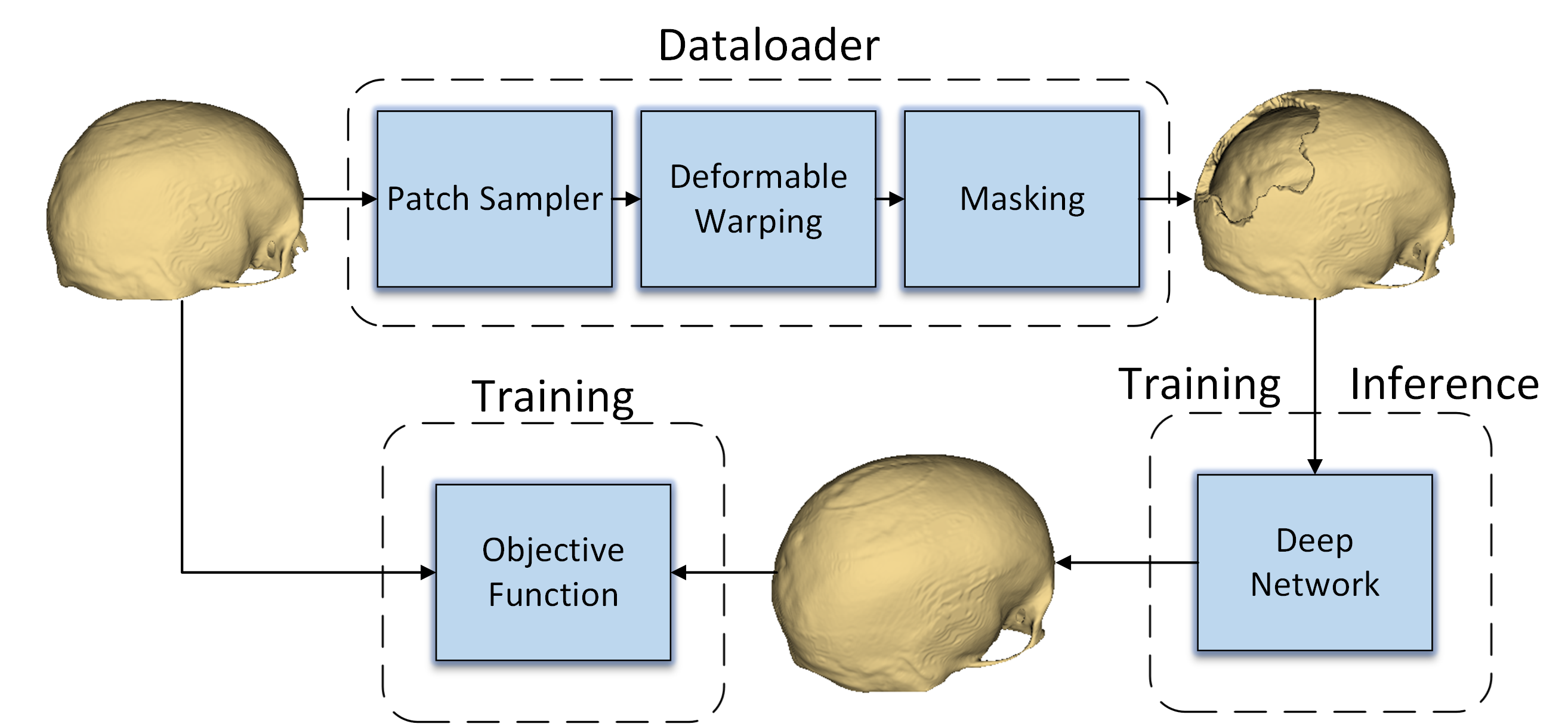
Automatic Cranial Defect Reconstruction with Self-Supervised Deep Deformable Masked Autoencoders
Marek Wodzinski, Daria Hemmerling, Mateusz Daniol
Thousands of people suffer from cranial injuries every year. They require personalized implants that need to be designed and manufactured before the reconstruction surgery. The manual design is expensive and time-consuming leading to searching for algorithms whose goal is to automatize the process. The problem can be formulated as volumetric shape completion and solved by deep neural networks dedicated to supervised image segmentation. However, such an approach requires annotating the ground-truth defects which is costly and time-consuming. Usually, the process is replaced with synthetic defect generation. However, even the synthetic ground-truth generation is time-consuming and limits the data heterogeneity, thus the deep models' generalizability. In our work, we propose an alternative and simple approach to use a self-supervised masked autoencoder to solve the problem. This approach by design increases the heterogeneity of the training set and can be seen as a form of data augmentation. We compare the proposed method with several state-of-the-art deep neural networks and show both the quantitative and qualitative improvement on the SkullBreak and SkullFix datasets. The proposed method can be used to efficiently reconstruct the cranial defects in real time.
Read more6/4/2024

0
SSAD: Self-supervised Auxiliary Detection Framework for Panoramic X-ray based Dental Disease Diagnosis
Zijian Cai, Xinquan Yang, Xuguang Li, Xiaoling Luo, Xuechen Li, Linlin Shen, He Meng, Yongqiang Deng
Panoramic X-ray is a simple and effective tool for diagnosing dental diseases in clinical practice. When deep learning models are developed to assist dentist in interpreting panoramic X-rays, most of their performance suffers from the limited annotated data, which requires dentist's expertise and a lot of time cost. Although self-supervised learning (SSL) has been proposed to address this challenge, the two-stage process of pretraining and fine-tuning requires even more training time and computational resources. In this paper, we present a self-supervised auxiliary detection (SSAD) framework, which is plug-and-play and compatible with any detectors. It consists of a reconstruction branch and a detection branch. Both branches are trained simultaneously, sharing the same encoder, without the need for finetuning. The reconstruction branch learns to restore the tooth texture of healthy or diseased teeth, while the detection branch utilizes these learned features for diagnosis. To enhance the encoder's ability to capture fine-grained features, we incorporate the image encoder of SAM to construct a texture consistency (TC) loss, which extracts image embedding from the input and output of reconstruction branch, and then enforces both embedding into the same feature space. Extensive experiments on the public DENTEX dataset through three detection tasks demonstrate that the proposed SSAD framework achieves state-of-the-art performance compared to mainstream object detection methods and SSL methods. The code is available at https://github.com/Dylonsword/SSAD
Read more6/21/2024