Semi-supervised variational autoencoder for cell feature extraction in multiplexed immunofluorescence images

0

Sign in to get full access
This summary was produced with help from an AI and may contain inaccuracies - check out the links to read the original source documents!
Related Papers
0
Semi-supervised variational autoencoder for cell feature extraction in multiplexed immunofluorescence images
Piumi Sandarenu, Julia Chen, Iveta Slapetova, Lois Browne, Peter H. Graham, Alexander Swarbrick, Ewan K. A. Millar, Yang Song, Erik Meijering
Advancements in digital imaging technologies have sparked increased interest in using multiplexed immunofluorescence (mIF) images to visualise and identify the interactions between specific immunophenotypes with the tumour microenvironment at the cellular level. Current state-of-the-art multiplexed immunofluorescence image analysis pipelines depend on cell feature representations characterised by morphological and stain intensity-based metrics generated using simple statistical and machine learning-based tools. However, these methods are not capable of generating complex representations of cells. We propose a deep learning-based cell feature extraction model using a variational autoencoder with supervision using a latent subspace to extract cell features in mIF images. We perform cell phenotype classification using a cohort of more than 44,000 multiplexed immunofluorescence cell image patches extracted across 1,093 tissue microarray cores of breast cancer patients, to demonstrate the success of our model against current and alternative methods.
Read more7/1/2024

0
Mew: Multiplexed Immunofluorescence Image Analysis through an Efficient Multiplex Network
Sukwon Yun, Jie Peng, Alexandro E. Trevino, Chanyoung Park, Tianlong Chen
Recent advancements in graph-based approaches for multiplexed immunofluorescence (mIF) images have significantly propelled the field forward, offering deeper insights into patient-level phenotyping. However, current graph-based methodologies encounter two primary challenges: (1) Cellular Heterogeneity, where existing approaches fail to adequately address the inductive biases inherent in graphs, particularly the homophily characteristic observed in cellular connectivity and; (2) Scalability, where handling cellular graphs from high-dimensional images faces difficulties in managing a high number of cells. To overcome these limitations, we introduce Mew, a novel framework designed to efficiently process mIF images through the lens of multiplex network. Mew innovatively constructs a multiplex network comprising two distinct layers: a Voronoi network for geometric information and a Cell-type network for capturing cell-wise homogeneity. This framework equips a scalable and efficient Graph Neural Network (GNN), capable of processing the entire graph during training. Furthermore, Mew integrates an interpretable attention module that autonomously identifies relevant layers for image classification. Extensive experiments on a real-world patient dataset from various institutions highlight Mew's remarkable efficacy and efficiency, marking a significant advancement in mIF image analysis. The source code of Mew can be found here: url{https://github.com/UNITES-Lab/Mew}
Read more7/26/2024
↗️
0
A Novel Generative Artificial Intelligence Method for Interference Study on Multiplex Brightfield Immunohistochemistry Images
Satarupa Mukherjee, Jim Martin, Yao Nie
Multiplex brightfield imaging offers the advantage of simultaneously analyzing multiple biomarkers on a single slide, as opposed to single biomarker labeling on multiple consecutive slides. To accurately analyze multiple biomarkers localized at the same cellular compartment, two representative biomarker sets were selected as assay models - cMET-PDL1-EGFR and CD8-LAG3-PDL1, where all three biomarkers can co-localize on the cell membrane. One of the most crucial preliminary stages for analyzing such assay is identifying each unique chromogen on individual cells. This is a challenging problem due to the co-localization of membrane stains from all the three biomarkers. It requires advanced color unmixing for creating the equivalent singleplex images from each triplex image for each biomarker. In this project, we developed a cycle-Generative Adversarial Network (cycle-GAN) method for unmixing the triplex images generated from the above-mentioned assays. Three different models were designed to generate the singleplex image for each of the three stains Tamra (purple), QM-Dabsyl (yellow) and Green. A notable novelty of our approach was that the input to the network were images in the optical density domain instead of conventionally used RGB images. The use of the optical density domain helped in reducing the blurriness of the synthetic singleplex images, which was often observed when the network was trained on RGB images. The cycle-GAN models were validated on 10,800 lung, gastric and colon images for the cMET-PDL1-EGFR assay and 3600 colon images for the CD8-LAG3-PDL1 assay. Visual as well as quantified assessments demonstrated that the proposed method is effective and efficient when compared with the manual reviewing results and is readily applicable to various multiplex assays.
Read more8/16/2024
0
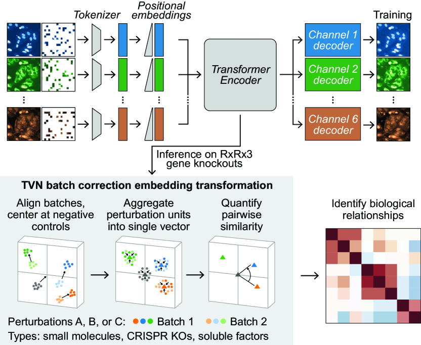
Masked Autoencoders for Microscopy are Scalable Learners of Cellular Biology
Oren Kraus, Kian Kenyon-Dean, Saber Saberian, Maryam Fallah, Peter McLean, Jess Leung, Vasudev Sharma, Ayla Khan, Jia Balakrishnan, Safiye Celik, Dominique Beaini, Maciej Sypetkowski, Chi Vicky Cheng, Kristen Morse, Maureen Makes, Ben Mabey, Berton Earnshaw
Featurizing microscopy images for use in biological research remains a significant challenge, especially for large-scale experiments spanning millions of images. This work explores the scaling properties of weakly supervised classifiers and self-supervised masked autoencoders (MAEs) when training with increasingly larger model backbones and microscopy datasets. Our results show that ViT-based MAEs outperform weakly supervised classifiers on a variety of tasks, achieving as much as a 11.5% relative improvement when recalling known biological relationships curated from public databases. Additionally, we develop a new channel-agnostic MAE architecture (CA-MAE) that allows for inputting images of different numbers and orders of channels at inference time. We demonstrate that CA-MAEs effectively generalize by inferring and evaluating on a microscopy image dataset (JUMP-CP) generated under different experimental conditions with a different channel structure than our pretraining data (RPI-93M). Our findings motivate continued research into scaling self-supervised learning on microscopy data in order to create powerful foundation models of cellular biology that have the potential to catalyze advancements in drug discovery and beyond.
Read more4/17/2024