Spectral U-Net: Enhancing Medical Image Segmentation via Spectral Decomposition

0

Sign in to get full access
Overview
- The paper presents a novel deep learning architecture called Spectral U-Net for medical image segmentation.
- Spectral U-Net leverages spectral decomposition to enhance the segmentation performance of the standard U-Net model.
- The key idea is to capture both spatial and spectral information in the network's feature representations.
Plain English Explanation
The goal of medical image segmentation is to automatically identify and outline different anatomical structures or regions of interest in medical scans like MRI or CT images. This is an important task in many clinical applications, such as diagnosis, treatment planning, and disease monitoring.
Spectral U-Net is a new deep learning model designed to improve the accuracy of medical image segmentation. It builds on the popular U-Net architecture, which has been widely used for this task, by incorporating a novel spectral decomposition component.
The key insight is that medical images not only have spatial information (the arrangement of pixels) but also spectral information (the frequency content of the image). Spectral U-Net aims to capture both of these types of information to create more robust and powerful feature representations for segmentation.
The spectral decomposition module breaks the input image into different frequency bands, similar to how the human visual system processes information. These spectral features are then combined with the spatial features learned by the standard U-Net layers to produce the final segmentation output.
By leveraging both spatial and spectral information, Spectral U-Net is able to outperform the standard U-Net and other state-of-the-art segmentation models on a range of medical imaging benchmarks. This suggests that incorporating spectral analysis can be a valuable addition to deep learning pipelines for medical image analysis.
Technical Explanation
The core of Spectral U-Net is the Dual Tree Complex Wavelet Transform (DT-CWT), which is used to decompose the input image into multiple frequency subbands. This allows the network to learn features at different scales and extract both low-level and high-level information from the image.
The DT-CWT module is integrated into the U-Net architecture, with the wavelet coefficients from each scale concatenated with the corresponding feature maps in the contracting and expansive paths of the U-Net. This enables the network to fuse the spatial and spectral representations at multiple levels of abstraction.
The authors also propose a novel Spectral Attention Module that selectively weights the different frequency subbands based on their relevance for the segmentation task. This helps the network focus on the most informative spectral features.
Extensive experiments on several medical image segmentation benchmarks, including cardiac, brain, and abdominal scans, demonstrate the effectiveness of Spectral U-Net. The model outperforms the standard U-Net and other state-of-the-art approaches, particularly in scenarios with limited training data or challenging anatomical structures.
Critical Analysis
One of the key strengths of Spectral U-Net is its ability to leverage both spatial and spectral information, which aligns well with the multi-scale nature of medical images. The authors provide a thorough evaluation across diverse datasets, showing the generalizability of their approach.
However, the paper does not delve into the interpretability of the learned spectral features or provide detailed analysis of how the different frequency subbands contribute to the segmentation performance. Further investigation into the internal workings of the model could yield additional insights.
Additionally, the computational overhead introduced by the spectral decomposition module may be a concern for real-time or resource-constrained applications. The authors mention the potential for further optimization, but this aspect could be explored in more depth.
It would also be interesting to see how Spectral U-Net performs on other types of medical imaging modalities, such as ultrasound or digital pathology, and whether the benefits generalize to these domains.
Conclusion
The Spectral U-Net architecture presented in this paper represents a promising advancement in medical image segmentation by seamlessly integrating spatial and spectral feature learning. The results demonstrate the value of incorporating spectral analysis into deep learning pipelines for medical image analysis, opening up new avenues for improving the accuracy and robustness of these critical clinical tools.
While the paper provides a solid technical foundation, further research could explore the interpretability of the learned spectral features, investigate computational efficiency, and evaluate the approach on a broader range of medical imaging modalities. Overall, Spectral U-Net stands as an innovative contribution to the field of medical image segmentation.
This summary was produced with help from an AI and may contain inaccuracies - check out the links to read the original source documents!
Related Papers
0
New!Spectral U-Net: Enhancing Medical Image Segmentation via Spectral Decomposition
Yaopeng Peng, Milan Sonka, Danny Z. Chen
This paper introduces Spectral U-Net, a novel deep learning network based on spectral decomposition, by exploiting Dual Tree Complex Wavelet Transform (DTCWT) for down-sampling and inverse Dual Tree Complex Wavelet Transform (iDTCWT) for up-sampling. We devise the corresponding Wave-Block and iWave-Block, integrated into the U-Net architecture, aiming at mitigating information loss during down-sampling and enhancing detail reconstruction during up-sampling. In the encoder, we first decompose the feature map into high and low-frequency components using DTCWT, enabling down-sampling while mitigating information loss. In the decoder, we utilize iDTCWT to reconstruct higher-resolution feature maps from down-sampled features. Evaluations on the Retina Fluid, Brain Tumor, and Liver Tumor segmentation datasets with the nnU-Net framework demonstrate the superiority of the proposed Spectral U-Net.
Read more9/17/2024
0
Harmonized Spatial and Spectral Learning for Robust and Generalized Medical Image Segmentation
Vandan Gorade, Sparsh Mittal, Debesh Jha, Rekha Singhal, Ulas Bagci
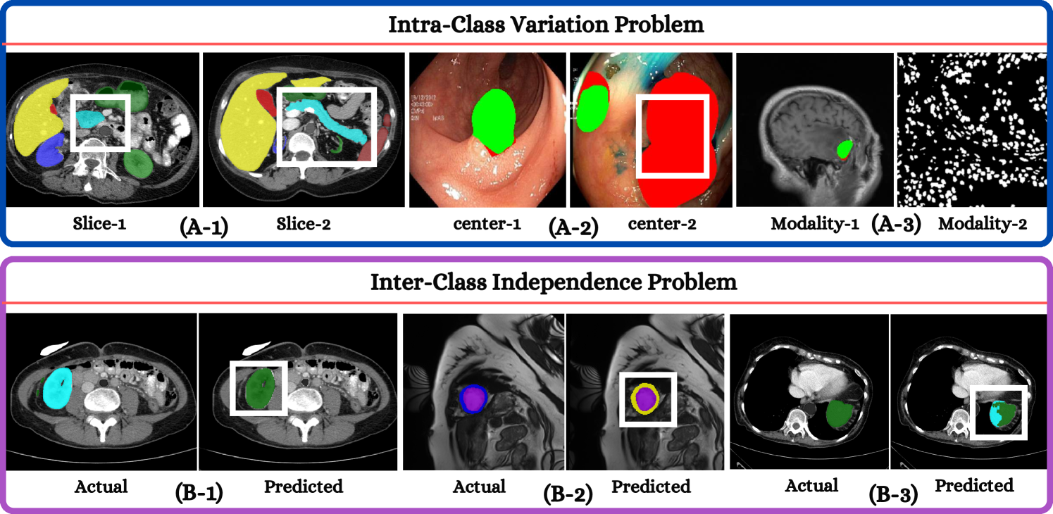
Deep learning has demonstrated remarkable achievements in medical image segmentation. However, prevailing deep learning models struggle with poor generalization due to (i) intra-class variations, where the same class appears differently in different samples, and (ii) inter-class independence, resulting in difficulties capturing intricate relationships between distinct objects, leading to higher false negative cases. This paper presents a novel approach that synergies spatial and spectral representations to enhance domain-generalized medical image segmentation. We introduce the innovative Spectral Correlation Coefficient objective to improve the model's capacity to capture middle-order features and contextual long-range dependencies. This objective complements traditional spatial objectives by incorporating valuable spectral information. Extensive experiments reveal that optimizing this objective with existing architectures like UNet and TransUNet significantly enhances generalization, interpretability, and noise robustness, producing more confident predictions. For instance, in cardiac segmentation, we observe a 0.81 pp and 1.63 pp (pp = percentage point) improvement in DSC over UNet and TransUNet, respectively. Our interpretability study demonstrates that, in most tasks, objectives optimized with UNet outperform even TransUNet by introducing global contextual information alongside local details. These findings underscore the versatility and effectiveness of our proposed method across diverse imaging modalities and medical domains.
Read more8/9/2024

0
WaveDH: Wavelet Sub-bands Guided ConvNet for Efficient Image Dehazing
Seongmin Hwang, Daeyoung Han, Cheolkon Jung, Moongu Jeon
The surge in interest regarding image dehazing has led to notable advancements in deep learning-based single image dehazing approaches, exhibiting impressive performance in recent studies. Despite these strides, many existing methods fall short in meeting the efficiency demands of practical applications. In this paper, we introduce WaveDH, a novel and compact ConvNet designed to address this efficiency gap in image dehazing. Our WaveDH leverages wavelet sub-bands for guided up-and-downsampling and frequency-aware feature refinement. The key idea lies in utilizing wavelet decomposition to extract low-and-high frequency components from feature levels, allowing for faster processing while upholding high-quality reconstruction. The downsampling block employs a novel squeeze-and-attention scheme to optimize the feature downsampling process in a structurally compact manner through wavelet domain learning, preserving discriminative features while discarding noise components. In our upsampling block, we introduce a dual-upsample and fusion mechanism to enhance high-frequency component awareness, aiding in the reconstruction of high-frequency details. Departing from conventional dehazing methods that treat low-and-high frequency components equally, our feature refinement block strategically processes features with a frequency-aware approach. By employing a coarse-to-fine methodology, it not only refines the details at frequency levels but also significantly optimizes computational costs. The refinement is performed in a maximum 8x downsampled feature space, striking a favorable efficiency-vs-accuracy trade-off. Extensive experiments demonstrate that our method, WaveDH, outperforms many state-of-the-art methods on several image dehazing benchmarks with significantly reduced computational costs. Our code is available at https://github.com/AwesomeHwang/WaveDH.
Read more4/3/2024

0
New!Unsupervised Hyperspectral and Multispectral Image Blind Fusion Based on Deep Tucker Decomposition Network with Spatial-Spectral Manifold Learning
He Wang, Yang Xu, Zebin Wu, Zhihui Wei
Hyperspectral and multispectral image fusion aims to generate high spectral and spatial resolution hyperspectral images (HR-HSI) by fusing high-resolution multispectral images (HR-MSI) and low-resolution hyperspectral images (LR-HSI). However, existing fusion methods encounter challenges such as unknown degradation parameters, incomplete exploitation of the correlation between high-dimensional structures and deep image features. To overcome these issues, in this article, an unsupervised blind fusion method for hyperspectral and multispectral images based on Tucker decomposition and spatial spectral manifold learning (DTDNML) is proposed. We design a novel deep Tucker decomposition network that maps LR-HSI and HR-MSI into a consistent feature space, achieving reconstruction through decoders with shared parameter. To better exploit and fuse spatial-spectral features in the data, we design a core tensor fusion network that incorporates a spatial spectral attention mechanism for aligning and fusing features at different scales. Furthermore, to enhance the capacity in capturing global information, a Laplacian-based spatial-spectral manifold constraints is introduced in shared-decoders. Sufficient experiments have validated that this method enhances the accuracy and efficiency of hyperspectral and multispectral fusion on different remote sensing datasets. The source code is available at https://github.com/Shawn-H-Wang/DTDNML.
Read more9/17/2024