Supporting Mitosis Detection AI Training with Inter-Observer Eye-Gaze Consistencies

0

Sign in to get full access
Overview
- The paper explores using eye-gaze tracking data to support training of AI systems for detecting mitosis (cell division) in pathology images.
- The researchers hypothesize that consistent patterns in how pathologists visually examine samples can provide useful training signals for mitosis detection models.
- The study involves collecting eye-gaze data from multiple pathologists examining the same pathology slides and analyzing the consistency in their visual attention patterns.
- The consistent eye-gaze patterns are then incorporated as additional training signals for a convolutional neural network model for mitosis detection.
Plain English Explanation
This research aims to help improve the performance of AI systems that can automatically detect cell division (mitosis) in microscope images of tissue samples. Accurately detecting mitotic cells is an important task in pathology, as it can provide insights into the growth and proliferation of cancer cells.
The key insight behind this work is that human experts, like pathologists, tend to exhibit consistent patterns in how they visually examine and analyze tissue samples under a microscope. By tracking and analyzing the eye movements of multiple pathologists looking at the same slides, the researchers found that there are reliable similarities in where the pathologists focus their attention.
The researchers hypothesized that these consistent eye-gaze patterns could serve as valuable training data to help an AI system learn to detect mitotic cells more accurately. So they incorporated the eye-gaze consistency information as an additional signal when training a convolutional neural network model for mitosis detection.
The idea is that by learning from the common visual scanning strategies of expert pathologists, the AI model can better recognize the telltale visual features and locations associated with cell division. This could make the mitosis detection model more robust and accurate compared to training it solely on labeled example images.
Technical Explanation
The study involved collecting eye-gaze tracking data from 8 board-certified pathologists as they visually examined a set of 100 digitized histopathology slides containing breast cancer tissue samples. The slides included annotations marking the locations of mitotic cells, which were used as ground truth labels.
The researchers then analyzed the eye-gaze data to quantify the consistency in visual attention patterns across the pathologists. They calculated measures like the proportion of slides where each pixel location received consistent gaze fixations from multiple observers. These consistency maps were used to generate "attention heatmaps" highlighting the regions of the slides that received the most consistent visual focus from the experts.
Next, the researchers incorporated this eye-gaze consistency information when training a convolutional neural network (CNN) model for automated mitosis detection. Specifically, they used the attention heatmaps to weight the loss function, placing more emphasis on correctly classifying pixels in regions that received consistent visual focus from the pathologists.
The results showed that the CNN model trained with the eye-gaze consistency signals outperformed a baseline CNN model trained only on the labeled mitosis examples. The eye-gaze-augmented model achieved higher F1-scores on the mitosis detection task, indicating improved precision and recall.
Critical Analysis
The paper provides a compelling proof-of-concept for leveraging human expert visual attention patterns to enhance the training of AI models for medical image analysis tasks. The findings suggest that incorporating this type of behavioral data can indeed boost model performance beyond what is achievable through standard supervised learning on labeled example data alone.
However, the study has some notable limitations. The experiments were conducted on a relatively small dataset of 100 slides from a single tissue type (breast cancer). More extensive evaluation across a broader range of pathology domains would be needed to fully assess the generalizability of the approach.
Additionally, the paper does not provide much insight into the specific mechanisms by which the eye-gaze consistency information improves the mitosis detection performance. Further analysis of the trained model's behavior and learned visual features could shed light on the underlying reasons for the performance boost.
It would also be valuable to explore how the eye-gaze-augmented training approach compares to other strategies for incorporating human expert knowledge, such as weakly-supervised or interactive learning techniques. Understanding the relative merits of different ways to leverage human expertise could help guide the development of more effective AI-assisted pathology tools.
Conclusion
Overall, this research demonstrates a promising new direction for boosting the performance of AI-based medical image analysis systems. By learning from the consistent visual scanning strategies of expert human observers, the mitosis detection model was able to become more accurate and reliable. This kind of human-in-the-loop training approach could have broad applicability across a range of pathology and radiology tasks, helping to bridge the gap between the capabilities of AI and the expertise of medical professionals.
As AI systems become increasingly integrated into clinical workflows, techniques like the one described in this paper will be crucial for ensuring that the technology is optimized to complement and empower human experts, rather than replace them. Continued research in this area has the potential to yield powerful AI-assisted diagnostic tools that can enhance the speed, consistency, and accuracy of healthcare delivery.
This summary was produced with help from an AI and may contain inaccuracies - check out the links to read the original source documents!
Related Papers
0
Supporting Mitosis Detection AI Training with Inter-Observer Eye-Gaze Consistencies
Hongyan Gu, Zihan Yan, Ayesha Alvi, Brandon Day, Chunxu Yang, Zida Wu, Shino Magaki, Mohammad Haeri, Xiang 'Anthony' Chen
The expansion of artificial intelligence (AI) in pathology tasks has intensified the demand for doctors' annotations in AI development. However, collecting high-quality annotations from doctors is costly and time-consuming, creating a bottleneck in AI progress. This study investigates eye-tracking as a cost-effective technology to collect doctors' behavioral data for AI training with a focus on the pathology task of mitosis detection. One major challenge in using eye-gaze data is the low signal-to-noise ratio, which hinders the extraction of meaningful information. We tackled this by levering the properties of inter-observer eye-gaze consistencies and creating eye-gaze labels from consistent eye-fixations shared by a group of observers. Our study involved 14 non-medical participants, from whom we collected eye-gaze data and generated eye-gaze labels based on varying group sizes. We assessed the efficacy of such eye-gaze labels by training Convolutional Neural Networks (CNNs) and comparing their performance to those trained with ground truth annotations and a heuristic-based baseline. Results indicated that CNNs trained with our eye-gaze labels closely followed the performance of ground-truth-based CNNs, and significantly outperformed the baseline. Although primarily focused on mitosis, we envision that insights from this study can be generalized to other medical imaging tasks.
Read more4/3/2024
0
Weakly-supervised Medical Image Segmentation with Gaze Annotations
Yuan Zhong, Chenhui Tang, Yumeng Yang, Ruoxi Qi, Kang Zhou, Yuqi Gong, Pheng Ann Heng, Janet H. Hsiao, Qi Dou
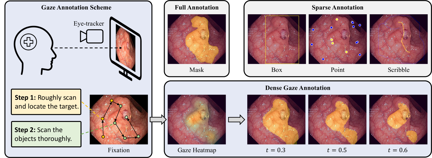
Eye gaze that reveals human observational patterns has increasingly been incorporated into solutions for vision tasks. Despite recent explorations on leveraging gaze to aid deep networks, few studies exploit gaze as an efficient annotation approach for medical image segmentation which typically entails heavy annotating costs. In this paper, we propose to collect dense weak supervision for medical image segmentation with a gaze annotation scheme. To train with gaze, we propose a multi-level framework that trains multiple networks from discriminative human attention, simulated with a set of pseudo-masks derived by applying hierarchical thresholds on gaze heatmaps. Furthermore, to mitigate gaze noise, a cross-level consistency is exploited to regularize overfitting noisy labels, steering models toward clean patterns learned by peer networks. The proposed method is validated on two public medical datasets of polyp and prostate segmentation tasks. We contribute a high-quality gaze dataset entitled GazeMedSeg as an extension to the popular medical segmentation datasets. To the best of our knowledge, this is the first gaze dataset for medical image segmentation. Our experiments demonstrate that gaze annotation outperforms previous label-efficient annotation schemes in terms of both performance and annotation time. Our collected gaze data and code are available at: https://github.com/med-air/GazeMedSeg.
Read more7/11/2024
🛸
0
Using artificial intelligence methods for the studyed visual analyzer
A. I. Medvedeva, M. V. Kholod
The paper describes how various techniques for applying artificial intelligence to the study of human eyes are utilized. The first dataset was collected using computerized perimetry to investigate the visualization of the human visual field and the diagnosis of glaucoma. A method to analyze the image using software tools is proposed. The second dataset was obtained, as part of the implementation of a Russian-Swiss experiment to collect and analyze eye movement data using the Tobii Pro Glasses 3 device on VR video. Eye movements and focus on the recorded route of a virtual journey through the canton of Vaud were investigated. Methods are being developed to investigate the dependencies of eye pupil movements using mathematical modelling. VR-video users can use these studies in medicine to assess the course and deterioration of glaucoma patients and to study the mechanisms of attention to tourist attractions.
Read more5/1/2024

0
Learning Gaze-aware Compositional GAN
Nerea Aranjuelo, Siyu Huang, Ignacio Arganda-Carreras, Luis Unzueta, Oihana Otaegui, Hanspeter Pfister, Donglai Wei
Gaze-annotated facial data is crucial for training deep neural networks (DNNs) for gaze estimation. However, obtaining these data is labor-intensive and requires specialized equipment due to the challenge of accurately annotating the gaze direction of a subject. In this work, we present a generative framework to create annotated gaze data by leveraging the benefits of labeled and unlabeled data sources. We propose a Gaze-aware Compositional GAN that learns to generate annotated facial images from a limited labeled dataset. Then we transfer this model to an unlabeled data domain to take advantage of the diversity it provides. Experiments demonstrate our approach's effectiveness in generating within-domain image augmentations in the ETH-XGaze dataset and cross-domain augmentations in the CelebAMask-HQ dataset domain for gaze estimation DNN training. We also show additional applications of our work, which include facial image editing and gaze redirection.
Read more6/3/2024