A systematic review: Deep learning-based methods for pneumonia region detection

0
🤿
Sign in to get full access
Overview
- Pneumonia is a leading cause of death worldwide, especially among children and adults.
- In the past 10 years, computer-aided methods using deep learning have been developed to improve pneumonia diagnosis.
- Deep learning has outperformed traditional machine learning approaches in detecting pneumonia.
- This review paper examines the mainstream deep learning methods used for pneumonia detection.
Plain English Explanation
Pneumonia is a serious lung infection that can be life-threatening, especially for young children and the elderly. In recent years, researchers have been working on developing computer-based tools to help doctors diagnose pneumonia more quickly and accurately. These tools use a type of artificial intelligence called deep learning, which has proven to be more effective than older machine learning methods at identifying signs of pneumonia in medical images.
This review paper looks at the different deep learning approaches that have been used for pneumonia detection. It covers the key details of these approaches, including the datasets they used, how the data was processed, the overall workflow, the results they achieved, their advantages, and their limitations. The paper also discusses the current challenges in this area of research and proposes ideas for future work that could further improve the performance of deep learning models in detecting, classifying, and pinpointing regions of the lungs affected by pneumonia.
The goal of this review is to provide a comprehensive summary and analysis of the current state of deep learning for pneumonia diagnosis. This can help guide future research and development efforts in this important field, with the ultimate aim of getting better tools into the hands of doctors to treat this deadly disease.
Technical Explanation
This review paper examines the use of deep learning approaches for the detection of pneumonia from medical images. The researchers searched the literature and identified the mainstream deep learning methods that have been applied to this problem.
The paper covers key aspects of these deep learning approaches, including the datasets used, data preprocessing and augmentation techniques, the overall workflow and architectures employed, the outcomes achieved, the advantages conferred by deep learning, and the limitations of the current methods.
The review also discusses the current challenges in this field of research and proposes potential future directions to enhance the research procedures and improve the overall performance of deep learning models in detecting, classifying, and localizing regions affected by pneumonia.
Critical Analysis
The review provides a comprehensive overview of the state-of-the-art deep learning approaches for pneumonia detection, highlighting their strengths and limitations. However, the paper does not delve into a detailed critique of the individual studies or the field as a whole.
For example, the review could have discussed the potential biases or limitations in the datasets used, which can impact the generalizability of the deep learning models. Additionally, the review could have explored the trade-offs between the different deep learning architectures and their suitability for real-world clinical deployment.
Furthermore, the review could have challenged some of the assumptions or design choices made in the reviewed studies, and suggested alternative directions for future research that could address the identified limitations. A more critical and forward-looking analysis would have strengthened the review and provided readers with a deeper understanding of the current state and future prospects of deep learning for pneumonia detection.
Conclusion
This review paper provides a comprehensive summary and analysis of the mainstream deep learning approaches that have been developed for the detection of pneumonia from medical images. The paper highlights the key aspects of these approaches, including their datasets, data processing techniques, architectures, and outcomes, as well as their advantages and limitations.
The review also discusses the current challenges in this field of research and proposes future directions to enhance the research procedures and improve the overall performance of deep learning models in detecting, classifying, and localizing regions affected by pneumonia. By offering this insightful summary, the review aims to facilitate the further development of deep learning approaches in addressing this treatable and potentially deadly disease.
This summary was produced with help from an AI and may contain inaccuracies - check out the links to read the original source documents!
Related Papers
🤿
0
A systematic review: Deep learning-based methods for pneumonia region detection
Xinmei Xu
Pneumonia disease is one of the leading causes of death among children and adults worldwide. In the last ten years, computer-aided pneumonia detection methods have been developed to improve the efficiency and accuracy of the diagnosis process. Among those methods, the effects of deep learning approaches surpassed that of other traditional machine learning methods. This review paper searched and examined existing mainstream deep-learning approaches in the detection of pneumonia regions. This paper focuses on key aspects of the collected research, including their datasets, data processing techniques, general workflow, outcomes, advantages, and limitations. This paper also discusses current challenges in the field and proposes future work that can be done to enhance research procedures and the overall performance of deep learning models in detecting, classifying, and localizing infected regions. This review aims to offer an insightful summary and analysis of current research, facilitating the development of deep learning approaches in addressing treatable diseases.
Read more8/27/2024
🤿
0
Pneumonia Diagnosis through pixels -- A Deep Learning Model for detection and classification
Amit Karanth Gurpur, Janani S, Ajeetha B, Brintha Therese A, Rajeswaran Rangasami
Manual identification and classification of pneumonia and COVID-19 infection is a cumbersome process that, if delayed can cause irreversible damage to the patient. We have compiled CT scan images from various sources, namely, from the China Consortium of Chest CT Image Investigation (CC-CCII), the Negin Radiology located at Sari in Iran, an open access COVID-19 repository from Havard dataverse, and Sri Ramachandra University, Chennai, India. The images were preprocessed using various methods such as normalization, sharpening, median filter application, binarizing, and cropping to ensure uniformity while training the models. We present an ensemble classification approach using deep learning and machine learning methods to classify patients with the said diseases. Our ensemble model uses pre-trained networks such as ResNet-18 and ResNet-50 for classification and MobileNetV2 for feature extraction. The features from MobileNetV2 are used by the gradient-boosting classifier for the classification of patients. Using ResNet-18, ResNet-50, and the MobileNetV2 aided gradient boosting classifier, we propose an ensemble model with an accuracy of 98 percent on unseen data.
Read more4/22/2024
0
Efficient and Accurate Pneumonia Detection Using a Novel Multi-Scale Transformer Approach
Alireza Saber, Pouria Parhami, Alimihammad Siahkarzadeh, Amirreza Fateh
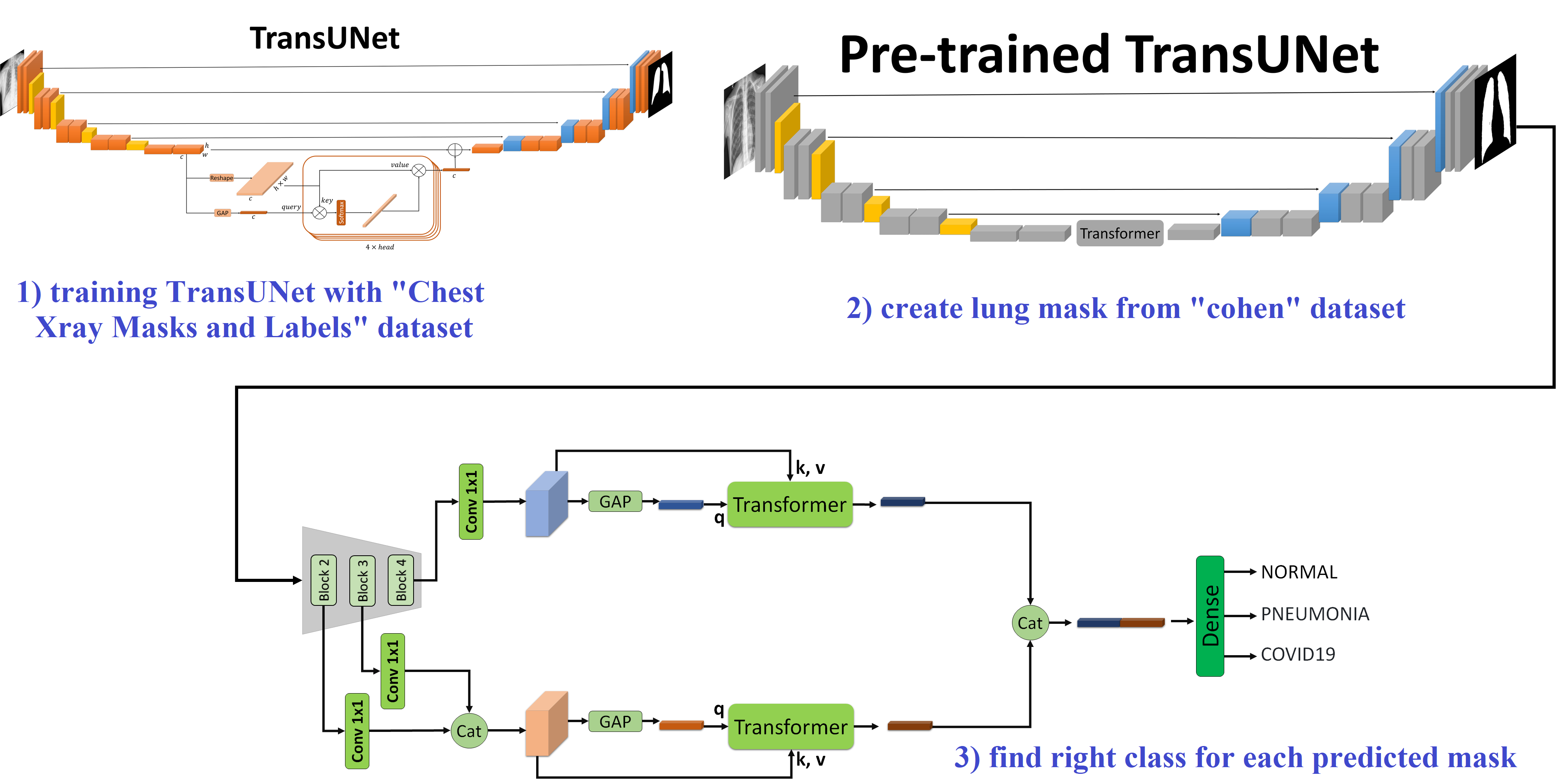
Pneumonia, a severe respiratory disease, poses significant diagnostic challenges, especially in underdeveloped regions. Traditional diagnostic methods, such as chest X-rays, suffer from variability in interpretation among radiologists, necessitating reliable automated tools. In this study, we propose a novel approach combining deep learning and transformer-based attention mechanisms to enhance pneumonia detection from chest X-rays. Our method begins with lung segmentation using a TransUNet model that integrates our specialized transformer module, which has fewer parameters compared to common transformers while maintaining performance. This model is trained on the Chest Xray Masks and Labels dataset and then applied to the Kermany and Cohen datasets to isolate lung regions, enhancing subsequent classification tasks. For classification, we employ pre-trained ResNet models (ResNet-50 and ResNet-101) to extract multi-scale feature maps, processed through our modified transformer module. By employing our specialized transformer, we attain superior results with significantly fewer parameters compared to common transformer models. Our approach achieves high accuracy rates of 92.79% on the Kermany dataset and 95.11% on the Cohen dataset, ensuring robust and efficient performance suitable for resource-constrained environments. https://github.com/amirrezafateh/Multi-Scale-Transformer-Pneumonia
Read more8/9/2024

0
InfLocNet: Enhanced Lung Infection Localization and Disease Detection from Chest X-Ray Images Using Lightweight Deep Learning
Md. Asiful Islam Miah, Shourin Paul, Sunanda Das, M. M. A. Hashem
In recent years, the integration of deep learning techniques into medical imaging has revolutionized the diagnosis and treatment of lung diseases, particularly in the context of COVID-19 and pneumonia. This paper presents a novel, lightweight deep learning based segmentation-classification network designed to enhance the detection and localization of lung infections using chest X-ray images. By leveraging the power of transfer learning with pre-trained VGG-16 weights, our model achieves robust performance even with limited training data. The architecture incorporates refined skip connections within the UNet++ framework, reducing semantic gaps and improving precision in segmentation tasks. Additionally, a classification module is integrated at the end of the encoder block, enabling simultaneous classification and segmentation. This dual functionality enhances the model's versatility, providing comprehensive diagnostic insights while optimizing computational efficiency. Experimental results demonstrate that our proposed lightweight network outperforms existing methods in terms of accuracy and computational requirements, making it a viable solution for real-time and resource constrained medical imaging applications. Furthermore, the streamlined design facilitates easier hyperparameter tuning and deployment on edge devices. This work underscores the potential of advanced deep learning architectures in improving clinical outcomes through precise and efficient medical image analysis. Our model achieved remarkable results with an Intersection over Union (IoU) of 93.59% and a Dice Similarity Coefficient (DSC) of 97.61% in lung area segmentation, and an IoU of 97.67% and a DSC of 87.61% for infection region localization. Additionally, it demonstrated high accuracy of 93.86% and sensitivity of 89.55% in detecting chest diseases, highlighting its efficacy and reliability.
Read more8/14/2024