TBConvL-Net: A Hybrid Deep Learning Architecture for Robust Medical Image Segmentation

0

Sign in to get full access
Overview
- This paper presents a new deep learning architecture called TBConvL-Net for robust medical image segmentation.
- It combines convolutional neural networks (CNNs), long short-term memory (LSTMs), and vision transformers to leverage the strengths of each approach.
- The goal is to improve the accuracy and robustness of medical image segmentation, which is crucial for various clinical applications.
Plain English Explanation
The paper introduces a new deep learning architecture called TBConvL-Net that is designed to tackle the challenge of medical image segmentation. Medical image segmentation is the process of dividing an image into different regions, such as organs or tumors, to help doctors better understand and diagnose medical conditions.
The TBConvL-Net architecture combines three powerful machine learning techniques:
-
Convolutional neural networks (CNNs): These are a type of deep learning model that excel at processing and understanding visual information, like medical images.
-
Long short-term memory (LSTMs): These are a type of recurrent neural network that can capture long-term dependencies in data, which can be useful for understanding the context and relationships within medical images.
-
Vision transformers: These are a more recent deep learning technique that can effectively model the global, long-range dependencies in visual data, which can complement the local and short-range processing of CNNs.
By combining these three techniques, the researchers aim to create a more accurate and robust medical image segmentation model that can handle the complexities and nuances of medical data. This could lead to better diagnostic tools and ultimately improved patient care.
Technical Explanation
The TBConvL-Net architecture consists of three main components:
-
Convolutional Neural Network (CNN) Encoder: This part of the model uses a CNN to extract visual features from the input medical images. The CNN encoder helps capture the local, spatial information in the images.
-
Long Short-Term Memory (LSTM) Decoder: This component uses an LSTM network to process the features extracted by the CNN encoder. The LSTM decoder can model the long-range dependencies and contextual information within the medical images, which is important for accurate segmentation.
-
Vision Transformer (ViT) Module: The ViT module is integrated into the architecture to capture the global, long-range relationships in the visual data. This complements the local and short-range processing of the CNN encoder.
The researchers trained and evaluated the TBConvL-Net model on several medical image segmentation tasks, including brain tumor, liver, and pancreas segmentation. They compared the performance of TBConvL-Net to other state-of-the-art models and found that it achieved superior results in terms of segmentation accuracy and robustness.
Critical Analysis
The paper presents a well-designed and thorough evaluation of the TBConvL-Net architecture. The researchers acknowledged some limitations, such as the need for further optimization of the model's hyperparameters and the potential for overfitting on certain datasets.
One area for further research could be investigating the interpretability and explainability of the TBConvL-Net model. Understanding the reasoning behind the model's predictions could be crucial for building trust and adoption in the medical community.
Additionally, the researchers could explore the generalization capabilities of TBConvL-Net by testing it on a wider range of medical imaging modalities and clinical applications. Demonstrating the model's ability to adapt to different medical contexts would strengthen the case for its real-world deployment.
Conclusion
The TBConvL-Net architecture presented in this paper represents a promising step forward in the field of medical image segmentation. By combining the strengths of CNNs, LSTMs, and vision transformers, the model achieves state-of-the-art performance and improved robustness compared to existing approaches.
This research could have significant implications for the development of more accurate and reliable medical imaging tools, which could ultimately lead to better patient outcomes and more efficient healthcare delivery. As the researchers continue to refine and expand the capabilities of TBConvL-Net, it could become a valuable asset in the arsenal of medical professionals tasked with early disease detection and monitoring.
This summary was produced with help from an AI and may contain inaccuracies - check out the links to read the original source documents!
Related Papers
0
TBConvL-Net: A Hybrid Deep Learning Architecture for Robust Medical Image Segmentation
Shahzaib Iqbal, Tariq M. Khan, Syed S. Naqvi, Asim Naveed, Erik Meijering
Deep learning has shown great potential for automated medical image segmentation to improve the precision and speed of disease diagnostics. However, the task presents significant difficulties due to variations in the scale, shape, texture, and contrast of the pathologies. Traditional convolutional neural network (CNN) models have certain limitations when it comes to effectively modelling multiscale context information and facilitating information interaction between skip connections across levels. To overcome these limitations, a novel deep learning architecture is introduced for medical image segmentation, taking advantage of CNNs and vision transformers. Our proposed model, named TBConvL-Net, involves a hybrid network that combines the local features of a CNN encoder-decoder architecture with long-range and temporal dependencies using biconvolutional long-short-term memory (LSTM) networks and vision transformers (ViT). This enables the model to capture contextual channel relationships in the data and account for the uncertainty of segmentation over time. Additionally, we introduce a novel composite loss function that considers both the segmentation robustness and the boundary agreement of the predicted output with the gold standard. Our proposed model shows consistent improvement over the state of the art on ten publicly available datasets of seven different medical imaging modalities.
Read more9/6/2024
0
D-TrAttUnet: Toward Hybrid CNN-Transformer Architecture for Generic and Subtle Segmentation in Medical Images
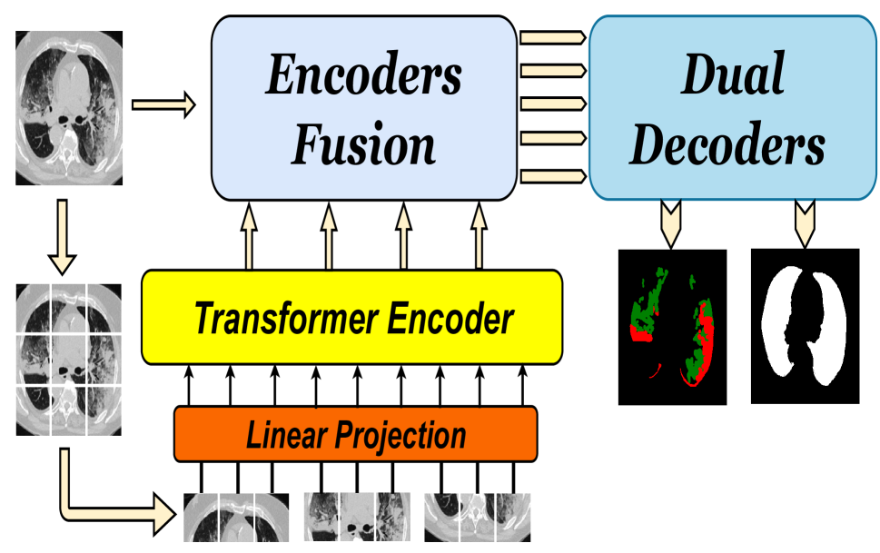
Fares Bougourzi, Fadi Dornaika, Cosimo Distante, Abdelmalik Taleb-Ahmed
Over the past two decades, machine analysis of medical imaging has advanced rapidly, opening up significant potential for several important medical applications. As complicated diseases increase and the number of cases rises, the role of machine-based imaging analysis has become indispensable. It serves as both a tool and an assistant to medical experts, providing valuable insights and guidance. A particularly challenging task in this area is lesion segmentation, a task that is challenging even for experienced radiologists. The complexity of this task highlights the urgent need for robust machine learning approaches to support medical staff. In response, we present our novel solution: the D-TrAttUnet architecture. This framework is based on the observation that different diseases often target specific organs. Our architecture includes an encoder-decoder structure with a composite Transformer-CNN encoder and dual decoders. The encoder includes two paths: the Transformer path and the Encoders Fusion Module path. The Dual-Decoder configuration uses two identical decoders, each with attention gates. This allows the model to simultaneously segment lesions and organs and integrate their segmentation losses. To validate our approach, we performed evaluations on the Covid-19 and Bone Metastasis segmentation tasks. We also investigated the adaptability of the model by testing it without the second decoder in the segmentation of glands and nuclei. The results confirmed the superiority of our approach, especially in Covid-19 infections and the segmentation of bone metastases. In addition, the hybrid encoder showed exceptional performance in the segmentation of glands and nuclei, solidifying its role in modern medical image analysis.
Read more5/8/2024

0
TESL-Net: A Transformer-Enhanced CNN for Accurate Skin Lesion Segmentation
Shahzaib Iqbal, Muhammad Zeeshan, Mehwish Mehmood, Tariq M. Khan, Imran Razzak
Early detection of skin cancer relies on precise segmentation of dermoscopic images of skin lesions. However, this task is challenging due to the irregular shape of the lesion, the lack of sharp borders, and the presence of artefacts such as marker colours and hair follicles. Recent methods for melanoma segmentation are U-Nets and fully connected networks (FCNs). As the depth of these neural network models increases, they can face issues like the vanishing gradient problem and parameter redundancy, potentially leading to a decrease in the Jaccard index of the segmentation model. In this study, we introduced a novel network named TESL-Net for the segmentation of skin lesions. The proposed TESL-Net involves a hybrid network that combines the local features of a CNN encoder-decoder architecture with long-range and temporal dependencies using bi-convolutional long-short-term memory (Bi-ConvLSTM) networks and a Swin transformer. This enables the model to account for the uncertainty of segmentation over time and capture contextual channel relationships in the data. We evaluated the efficacy of TESL-Net in three commonly used datasets (ISIC 2016, ISIC 2017, and ISIC 2018) for the segmentation of skin lesions. The proposed TESL-Net achieves state-of-the-art performance, as evidenced by a significantly elevated Jaccard index demonstrated by empirical results.
Read more8/20/2024

0
New!TTT-Unet: Enhancing U-Net with Test-Time Training Layers for biomedical image segmentation
Rong Zhou, Zhengqing Yuan, Zhiling Yan, Weixiang Sun, Kai Zhang, Yiwei Li, Yanfang Ye, Xiang Li, Lifang He, Lichao Sun
Biomedical image segmentation is crucial for accurately diagnosing and analyzing various diseases. However, Convolutional Neural Networks (CNNs) and Transformers, the most commonly used architectures for this task, struggle to effectively capture long-range dependencies due to the inherent locality of CNNs and the computational complexity of Transformers. To address this limitation, we introduce TTT-Unet, a novel framework that integrates Test-Time Training (TTT) layers into the traditional U-Net architecture for biomedical image segmentation. TTT-Unet dynamically adjusts model parameters during the testing time, enhancing the model's ability to capture both local and long-range features. We evaluate TTT-Unet on multiple medical imaging datasets, including 3D abdominal organ segmentation in CT and MR images, instrument segmentation in endoscopy images, and cell segmentation in microscopy images. The results demonstrate that TTT-Unet consistently outperforms state-of-the-art CNN-based and Transformer-based segmentation models across all tasks. The code is available at https://github.com/rongzhou7/TTT-Unet.
Read more9/20/2024