Towards Full Integration of Artificial Intelligence in Colon Capsule Endoscopy's Pathway

0
🤖
Sign in to get full access
Overview
- This study focuses on improving the integration of AI in colon capsule endoscopy (CCE) for the early detection and characterization of colorectal polyps.
- The researchers developed an AI-powered recognition network that can detect colorectal polyps with high sensitivity (99.9%), specificity (99.4%), and negative predictive value (99.8%).
- After polyp detection, separate networks were used to classify the polyps as neoplastic or non-neoplastic, and to estimate the size of the polyps with high accuracy.
- By automating these crucial image processing steps, the study aims to bring CCE closer to the capabilities of its counterpart, optical colonoscopy (OC), and enable its wider adoption in clinical practice.
Plain English Explanation
Colon capsule endoscopy (CCE) is a promising technology for early diagnosis of colorectal diseases, but it has not yet reached the same level of capability as traditional optical colonoscopy (OC). This study sought to address this gap by integrating advanced AI algorithms into the CCE process.
The researchers developed a specialized AI model that can automatically detect the presence of polyps (abnormal growths) in the colon with an extremely high degree of accuracy. This model was able to identify polyps with 99.9% sensitivity, meaning it rarely missed any polyps that were present. It also had a 99.4% specificity, indicating it could reliably distinguish polyps from other normal structures.
After the AI model detected a polyp, the researchers used two additional AI networks to analyze the characteristics of the polyp. One network could classify the polyp as either "neoplastic" (precancerous) or "non-neoplastic" with 82% sensitivity and 80% specificity. The other network could accurately measure the size of the polyp, getting it right 88% of the time.
By automating these critical image analysis steps, the researchers have taken an important step towards integrating AI seamlessly into the CCE process. This could help make CCE a more viable alternative to traditional OC, potentially improving early detection of colorectal diseases and expanding access to these crucial screening procedures.
Technical Explanation
The researchers developed a multi-stage AI system to address the gap between the current capabilities of colon capsule endoscopy (CCE) and optical colonoscopy (OC). The core of their approach was a polyp detection network that could identify the presence of polyps in CCE images with high accuracy.
This polyp detection network used advanced deep learning techniques to analyze the CCE images. It achieved an impressive 99.9% sensitivity, 99.4% specificity, and 99.8% negative predictive value in identifying polyps. This means the network was able to catch nearly every polyp present, with very few false positive or false negative results.
After the polyp detection step, the researchers fed the identified polyp images into two parallel AI networks. One network focused on classifying the polyps as either neoplastic (precancerous) or non-neoplastic, reaching 82% sensitivity and 80% specificity. The other network aimed to accurately estimate the size of the polyps, achieving an 88% accuracy rate.
By seamlessly integrating these AI-powered detection, characterization, and measurement capabilities into the CCE workflow, the researchers have taken a significant step towards bridging the gap between CCE and the gold standard of OC. This could pave the way for wider adoption of CCE in clinical practice, improving the early diagnosis of colorectal diseases.
Critical Analysis
The researchers have made a compelling case for the integration of advanced AI into the colon capsule endoscopy (CCE) workflow. The high performance of their polyp detection network, as well as the ability to characterize and measure polyps, represents a substantial advancement in the field.
However, the paper does not address some potential limitations or areas for further research. For example, the study was conducted on a relatively small dataset, and it's unclear how the AI models would perform on a larger, more diverse set of CCE images. Additionally, the researchers did not provide detailed information on the generalizability of their approach, such as how it might perform on data from different institutions or with varying imaging equipment.
Further research could also explore the integration of AI-powered CCE with other diagnostic tools, such as optical colonoscopy or other imaging modalities, to create a more comprehensive and accurate colorectal disease screening and diagnostic pipeline.
Additionally, the researchers did not delve into the interpretability of their AI models, which could be an important consideration for their clinical adoption and acceptance. Providing healthcare professionals with a better understanding of how the AI systems arrive at their decisions could enhance trust and facilitate their integration into routine practice.
Conclusion
This study represents a significant advance in the integration of AI into the colon capsule endoscopy (CCE) workflow. By developing highly accurate AI models for polyp detection, characterization, and size estimation, the researchers have taken an important step towards bridging the gap between CCE and the gold standard of optical colonoscopy (OC).
If these AI-powered capabilities can be further validated and refined, it could lead to wider adoption of CCE as a screening and diagnostic tool for colorectal diseases. This, in turn, could improve early detection and management of these conditions, potentially leading to better patient outcomes and reduced healthcare costs.
However, the researchers should address the limitations identified in the critical analysis, such as exploring the generalizability of their approach and the interpretability of their AI models, to ensure the long-term clinical viability and acceptance of their innovations.
This summary was produced with help from an AI and may contain inaccuracies - check out the links to read the original source documents!
Related Papers
🤖
0
Towards Full Integration of Artificial Intelligence in Colon Capsule Endoscopy's Pathway
Esmaeil S. Nadimi, Jan-Matthias Braun, Benedicte Schelde-Olesen, Emile Prudhomme, Victoria Blanes-Vidal, Gunnar Baatrup
Despite recent surge of interest in deploying colon capsule endoscopy (CCE) for early diagnosis of colorectal diseases, there remains a large gap between the current state of CCE in clinical practice, and the state of its counterpart optical colonoscopy (OC). Our study is aimed at closing this gap, by focusing on the full integration of AI in CCE's pathway, where image processing steps linked to the detection, localization and characterisation of important findings are carried out autonomously using various AI algorithms. We developed a recognition network, that with an impressive sensitivity of 99.9%, a specificity of 99.4%, and a negative predictive value (NPV) of 99.8%, detected colorectal polyps. After recognising a polyp within a sequence of images, only those images containing polyps were fed into two parallel independent networks for characterisation, and estimation of the size of those important findings. The characterisation network reached a sensitivity of 82% and a specificity of 80% in classifying polyps to two groups, namely neoplastic vs. non-neoplastic. The size estimation network reached an accuracy of 88% in correctly segmenting the polyps. By automatically incorporating this crucial information into CCE's pathway, we moved a step closer towards the full integration of AI in CCE's routine clinical practice.
Read more6/17/2024

0
Artificial Intelligence in Gastrointestinal Bleeding Analysis for Video Capsule Endoscopy: Insights, Innovations, and Prospects (2008-2023)
Tanisha Singh, Shreshtha Jha, Nidhi Bhatt, Palak Handa, Nidhi Goel, Sreedevi Indu
The escalating global mortality and morbidity rates associated with gastrointestinal (GI) bleeding, compounded by the complexities and limitations of traditional endoscopic methods, underscore the urgent need for a critical review of current methodologies used for addressing this condition. With an estimated 300,000 annual deaths worldwide, the demand for innovative diagnostic and therapeutic strategies is paramount. The introduction of Video Capsule Endoscopy (VCE) has marked a significant advancement, offering a comprehensive, non-invasive visualization of the digestive tract that is pivotal for detecting bleeding sources unattainable by traditional methods. Despite its benefits, the efficacy of VCE is hindered by diagnostic challenges, including time-consuming analysis and susceptibility to human error. This backdrop sets the stage for exploring Machine Learning (ML) applications in automating GI bleeding detection within capsule endoscopy, aiming to enhance diagnostic accuracy, reduce manual labor, and improve patient outcomes. Through an exhaustive analysis of 113 papers published between 2008 and 2023, this review assesses the current state of ML methodologies in bleeding detection, highlighting their effectiveness, challenges, and prospective directions. It contributes an in-depth examination of AI techniques in VCE frame analysis, offering insights into open-source datasets, mathematical performance metrics, and technique categorization. The paper sets a foundation for future research to overcome existing challenges, advancing gastrointestinal diagnostics through interdisciplinary collaboration and innovation in ML applications.
Read more9/4/2024
👀
0
Validating polyp and instrument segmentation methods in colonoscopy through Medico 2020 and MedAI 2021 Challenges
Debesh Jha, Vanshali Sharma, Debapriya Banik, Debayan Bhattacharya, Kaushiki Roy, Steven A. Hicks, Nikhil Kumar Tomar, Vajira Thambawita, Adrian Krenzer, Ge-Peng Ji, Sahadev Poudel, George Batchkala, Saruar Alam, Awadelrahman M. A. Ahmed, Quoc-Huy Trinh, Zeshan Khan, Tien-Phat Nguyen, Shruti Shrestha, Sabari Nathan, Jeonghwan Gwak, Ritika K. Jha, Zheyuan Zhang, Alexander Schlaefer, Debotosh Bhattacharjee, M. K. Bhuyan, Pradip K. Das, Deng-Ping Fan, Sravanthi Parsa, Sharib Ali, Michael A. Riegler, P{aa}l Halvorsen, Thomas De Lange, Ulas Bagci
Automatic analysis of colonoscopy images has been an active field of research motivated by the importance of early detection of precancerous polyps. However, detecting polyps during the live examination can be challenging due to various factors such as variation of skills and experience among the endoscopists, lack of attentiveness, and fatigue leading to a high polyp miss-rate. Deep learning has emerged as a promising solution to this challenge as it can assist endoscopists in detecting and classifying overlooked polyps and abnormalities in real time. In addition to the algorithm's accuracy, transparency and interpretability are crucial to explaining the whys and hows of the algorithm's prediction. Further, most algorithms are developed in private data, closed source, or proprietary software, and methods lack reproducibility. Therefore, to promote the development of efficient and transparent methods, we have organized the Medico automatic polyp segmentation (Medico 2020) and MedAI: Transparency in Medical Image Segmentation (MedAI 2021) competitions. We present a comprehensive summary and analyze each contribution, highlight the strength of the best-performing methods, and discuss the possibility of clinical translations of such methods into the clinic. For the transparency task, a multi-disciplinary team, including expert gastroenterologists, accessed each submission and evaluated the team based on open-source practices, failure case analysis, ablation studies, usability and understandability of evaluations to gain a deeper understanding of the models' credibility for clinical deployment. Through the comprehensive analysis of the challenge, we not only highlight the advancements in polyp and surgical instrument segmentation but also encourage qualitative evaluation for building more transparent and understandable AI-based colonoscopy systems.
Read more5/8/2024
0
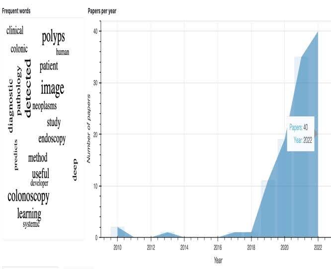
PubTrend: General Overview of Artificial Intelligence for Colorectal cancer diagnosis from 2010-2022
Mary Adewunmi, Reem Abdel-Salam
Colorectal cancer (CRC) is among the most prevalent cancers in the world. Due to numerous scholarly papers and broad enquiries about specific use cases for artificial intelligence (AI) in colorectal cancer, researchers find it challenging to explore relevant papers on the current knowledge, comprehensive knowledge, and past methodologies in the literature review. This review extracts recent AI technology advances for diagnosing colorectal cancer from January 2010 to March 2022. PubTrends was used to identify and automate the intellectual structure and comparable papers on the use of AI in colorectal cancer diagnosis using the most cited papers, keywords, and similar papers. Papers with quantitative results were represented with a tabular summary, and other paper contributions were in a sentence summary. Twenty-four (24) out of the forty-nine (49) top-cited papers were quantitative results, with one (1) outlier about lung cancer comprehensive screening. The most frequently used words were: polyps, detected, image, and colonoscopy. In addition, 83 per cent of the terms frequently used shortly before 2022 were image, polyps, detected, colonoscopy, and learning. In addition, 16 per cent are preparation, variant, classification, sample, and surgery. The review showcases 49 of the 50 most cited papers, their notable contributions, objectives, specific AI methods, results, conclusions, and further recommendations. These papers highlight the limitations of colonoscopy for therapeutic use. The review concluded that despite the enormous benefits of using artificial intelligence, from improving diagnosis, the medical AI programmer still needs to be actively involved in the diagnosis team for effective results in CRC diagnosis.
Read more7/10/2024