Transformer-based segmentation of adnexal lesions and ovarian implants in CT images

0
📉
Sign in to get full access
Overview
- Two transformer-based AI models, SMIT and Swin UNETR, were evaluated for their ability to accurately segment ovarian cancer tumors in CT images.
- The models performed better at segmenting tumors in the adnexa (the area around the ovaries) compared to omental implants (tumors on the omentum, a fatty apron-like structure in the abdomen).
- AI-assisted labeling of omental implants reduced the manual editing effort required compared to full manual correction.
- Both models had few false detections of tumors in the urinary bladder and small bowel.
Plain English Explanation
The researchers tested two advanced AI models, known as SMIT and Swin UNETR, on a dataset of CT scans from ovarian cancer patients. These models are trained to automatically identify and outline the boundaries of tumors in medical images.
The results showed that both SMIT and Swin UNETR were able to accurately segment the tumors, especially those located in the adnexa (the area around the ovaries). However, they had more difficulty accurately delineating the omental implants, which are tumors that have spread to the omentum, a fatty structure in the abdomen.
To address this, the researchers used a technique called AI-assisted labeling, where the models were used to partially label the omental implants. This reduced the amount of manual editing required to correct the labels, making the process more efficient.
Importantly, the models did not frequently mistake other structures, like the urinary bladder or small bowel, for tumors, which is a common problem with AI-based medical image analysis. This suggests the models are able to distinguish ovarian cancer tumors from other anatomical features with a high degree of accuracy.
Technical Explanation
The researchers fine-tuned two pre-trained transformer-based segmentation models, SMIT and Swin UNETR, on a dataset of CT images from ovarian cancer patients. Transformers are a type of deep learning architecture that has shown impressive performance in a variety of computer vision tasks.
The models were evaluated on an independent test set of CT scans. Both SMIT and Swin UNETR were able to accurately segment the tumors, particularly those located in the adnexa. However, they struggled more with the omental implants, which are often smaller and more diffuse.
To address this, the researchers used an AI-assisted labeling approach, where the models were used to partially label the omental implants. This reduced the manual editing effort required to correct the labels by over 60% compared to full manual correction. The AI-assisted labels also resulted in improved overall accuracy performance.
Importantly, the models did not generate many false positive detections in the urinary bladder and small bowel, which is a common challenge in medical image segmentation. On average, SMIT had 2.16 cc of false detections in these regions, while Swin UNETR had 7.37 cc.
Critical Analysis
The study provides a promising demonstration of the potential for transformer-based models to assist in the accurate segmentation of ovarian cancer tumors from CT scans. The researchers' use of AI-assisted labeling to improve the models' performance on challenging omental implants is a creative and practical approach.
However, the study is limited by its focus on a single dataset and tumor type. It would be valuable to see how the models perform on a more diverse set of medical imaging data, including other cancer types and anatomical regions. Additionally, the paper does not provide much insight into the inner workings of the models or the specific factors that contribute to their strengths and weaknesses.
Further research could also explore the clinical implications and potential real-world impact of these AI-powered segmentation tools. For example, how they might integrate with existing clinical workflows, the degree of time and effort they could save clinicians, and their effects on patient outcomes.
Overall, this study represents an important step forward in the application of advanced deep learning models to the challenging task of medical image analysis. By continuing to rigorously evaluate and improve these technologies, researchers can work towards realizing their potential to enhance clinical decision-making and patient care.
Conclusion
This research demonstrates the ability of two transformer-based AI models, SMIT and Swin UNETR, to accurately segment ovarian cancer tumors in CT scans. The models performed particularly well on tumors in the adnexa, and the use of AI-assisted labeling helped improve their performance on more challenging omental implants.
While further research is needed to fully understand the capabilities and limitations of these models, this study suggests they have the potential to assist clinicians in the diagnosis and management of ovarian cancer. By continuing to develop and refine advanced AI-based segmentation tools, researchers can work towards improving patient outcomes and reducing the burden on healthcare systems.
This summary was produced with help from an AI and may contain inaccuracies - check out the links to read the original source documents!
Related Papers
📉
0
Transformer-based segmentation of adnexal lesions and ovarian implants in CT images
Aneesh Rangnekar, Kevin M. Boehm, Emily A. Aherne, Ines Nikolovski, Natalie Gangai, Ying Liu, Dimitry Zamarin, Kara L. Roche, Sohrab P. Shah, Yulia Lakhman, Harini Veeraraghavan
Two self-supervised pretrained transformer-based segmentation models (SMIT and Swin UNETR) fine-tuned on a dataset of ovarian cancer CT images provided reasonably accurate delineations of the tumors in an independent test dataset. Tumors in the adnexa were segmented more accurately by both transformers (SMIT and Swin UNETR) than the omental implants. AI-assisted labeling performed on 72 out of 245 omental implants resulted in smaller manual editing effort of 39.55 mm compared to full manual correction of partial labels of 106.49 mm and resulted in overall improved accuracy performance. Both SMIT and Swin UNETR did not generate any false detection of omental metastases in the urinary bladder and relatively few false detections in the small bowel, with 2.16 cc on average for SMIT and 7.37 cc for Swin UNETR respectively.
Read more6/26/2024

0
Quantifying uncertainty in lung cancer segmentation with foundation models applied to mixed-domain datasets
Aneesh Rangnekar, Nishant Nadkarni, Jue Jiang, Harini Veeraraghavan
Medical image foundation models have shown the ability to segment organs and tumors with minimal fine-tuning. These models are typically evaluated on task-specific in-distribution (ID) datasets. However, reliable performance on ID dataset does not guarantee robust generalization on out-of-distribution (OOD) datasets. Importantly, once deployed for clinical use, it is impractical to have `ground truth' delineations to assess ongoing performance drifts, especially when images fall into OOD category due to different imaging protocols. Hence, we introduced a comprehensive set of computationally fast metrics to evaluate the performance of multiple foundation models (Swin UNETR, SimMIM, iBOT, SMIT) trained with self-supervised learning (SSL). SSL pretraining was selected as this approach is applicable for large, diverse, and unlabeled image sets. All models were fine-tuned on identical datasets for lung tumor segmentation from computed tomography (CT) scans. SimMIM, iBOT, and SMIT used identical architecture, pretraining, and fine-tuning datasets to assess performance variations with the choice of pretext tasks used in SSL. Evaluation was performed on two public lung cancer datasets (LRAD: n = 140, 5Rater: n = 21) with different image acquisitions and tumor stage compared to training data (n = 317 public resource with stage III-IV lung cancers) and a public non-cancer dataset containing volumetric CT scans of patients with pulmonary embolism (n = 120). All models produced similarly accurate tumor segmentation on the lung cancer testing datasets. SMIT produced a highest F1-score (LRAD: 0.60, 5Rater: 0.64) and lowest entropy (LRAD: 0.06, 5Rater: 0.12), indicating higher tumor detection rate and confident segmentations. In the OOD dataset, SMIT misdetected least number of tumors, indicated by median volume occupancy of 5.67 cc compared to second best method SimMIM of 9.97 cc.
Read more9/5/2024
0
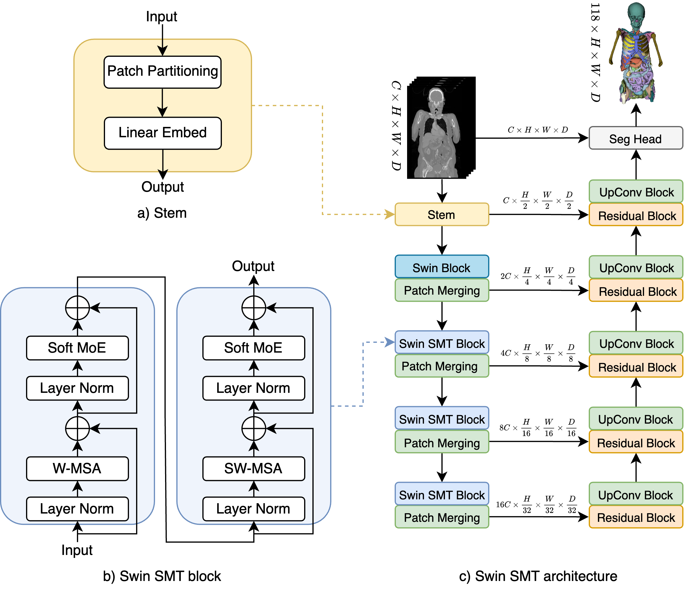
Swin SMT: Global Sequential Modeling in 3D Medical Image Segmentation
Szymon P{l}otka, Maciej Chrabaszcz, Przemyslaw Biecek
Recent advances in Vision Transformers (ViTs) have significantly enhanced medical image segmentation by facilitating the learning of global relationships. However, these methods face a notable challenge in capturing diverse local and global long-range sequential feature representations, particularly evident in whole-body CT (WBCT) scans. To overcome this limitation, we introduce Swin Soft Mixture Transformer (Swin SMT), a novel architecture based on Swin UNETR. This model incorporates a Soft Mixture-of-Experts (Soft MoE) to effectively handle complex and diverse long-range dependencies. The use of Soft MoE allows for scaling up model parameters maintaining a balance between computational complexity and segmentation performance in both training and inference modes. We evaluate Swin SMT on the publicly available TotalSegmentator-V2 dataset, which includes 117 major anatomical structures in WBCT images. Comprehensive experimental results demonstrate that Swin SMT outperforms several state-of-the-art methods in 3D anatomical structure segmentation, achieving an average Dice Similarity Coefficient of 85.09%. The code and pre-trained weights of Swin SMT are publicly available at https://github.com/MI2DataLab/SwinSMT.
Read more7/11/2024

0
Neuro-TransUNet: Segmentation of stroke lesion in MRI using transformers
Muhammad Nouman, Mohamed Mabrok, Essam A. Rashed
Accurate segmentation of the stroke lesions using magnetic resonance imaging (MRI) is associated with difficulties due to the complicated anatomy of the brain and the different properties of the lesions. This study introduces the Neuro-TransUNet framework, which synergizes the U-Net's spatial feature extraction with SwinUNETR's global contextual processing ability, further enhanced by advanced feature fusion and segmentation synthesis techniques. The comprehensive data pre-processing pipeline improves the framework's efficiency, which involves resampling, bias correction, and data standardization, enhancing data quality and consistency. Ablation studies confirm the significant impact of the advanced integration of U-Net with SwinUNETR and data pre-processing pipelines on performance and demonstrate the model's effectiveness. The proposed Neuro-TransUNet model, trained with the ATLAS v2.0 emph{training} dataset, outperforms existing deep learning algorithms and establishes a new benchmark in stroke lesion segmentation.
Read more6/11/2024