Tumor aware recurrent inter-patient deformable image registration of computed tomography scans with lung cancer

0
Sign in to get full access
Overview
- The paper presents a novel "tumor-aware recurrent inter-patient deformable image registration" approach for aligning computed tomography (CT) scans of patients with lung cancer.
- The method aims to accurately capture the deformation of lung tumors across different patient scans during the registration process.
- The approach uses a recurrent neural network architecture and incorporates tumor-specific information to guide the deformation.
Plain English Explanation
The paper describes a new way to align or match up different CT scans of lung cancer patients. The goal is to accurately capture how the lung tumors change shape and position across the different scans.
The key idea is to use a recurrent neural network, which is a type of machine learning model that can process information sequentially. This allows the model to consider the tumor's history and characteristics when aligning the scans.
By incorporating tumor-specific information, the method aims to ensure the deformation of the lung tumors is accurately captured during the alignment process. This is important because the tumors are the key structures of interest when analyzing these medical scans.
Technical Explanation
The paper presents a tumor-aware recurrent inter-patient deformable image registration approach for aligning CT scans of lung cancer patients. The method uses a recurrent neural network architecture to capture the deformation of lung tumors across different patient scans during the registration process.
The key components of the approach include:
-
Tumor-Aware Registration: The method incorporates tumor-specific information, such as the tumor's location, size, and shape, to guide the deformation process and ensure the tumors are accurately aligned across scans.
-
Recurrent Architecture: The use of a recurrent neural network allows the model to process the scans sequentially and consider the tumor's history and characteristics when aligning the images.
-
Inter-Patient Registration: The approach is designed to work across different patient scans, enabling the alignment of lung CT scans from multiple individuals with lung cancer.
By combining these elements, the proposed method aims to achieve more accurate and robust deformable image registration for lung cancer applications, where the precise alignment of tumors is crucial for tasks such as disease monitoring and treatment planning.
Critical Analysis
The paper presents a novel and potentially valuable approach for deformable image registration in the context of lung cancer CT scans. The incorporation of tumor-specific information and the use of a recurrent neural network architecture are interesting and well-motivated ideas.
However, the paper does not provide a comprehensive evaluation of the method's performance, particularly in comparison to other state-of-the-art deformable registration techniques. It would be helpful to see more extensive experiments and comparisons to better understand the method's strengths, limitations, and potential advantages over existing approaches.
Additionally, the paper does not address potential challenges or limitations of the proposed method, such as the availability and reliability of tumor segmentation information, the computational complexity of the recurrent architecture, or the generalization of the approach to other types of cancer or medical imaging modalities.
Further research and validation of the method on larger and more diverse datasets would be beneficial to assess its robustness and practical applicability in real-world clinical settings.
Conclusion
This paper introduces a novel "tumor-aware recurrent inter-patient deformable image registration" approach for aligning CT scans of lung cancer patients. The method incorporates tumor-specific information and uses a recurrent neural network architecture to accurately capture the deformation of lung tumors across different patient scans.
The proposed approach has the potential to improve the accuracy and robustness of deformable image registration in lung cancer applications, where the precise alignment of tumors is crucial for tasks such as disease monitoring and treatment planning. However, the paper would benefit from more comprehensive evaluation and discussion of the method's limitations and areas for further research.
This summary was produced with help from an AI and may contain inaccuracies - check out the links to read the original source documents!
Related Papers
0
New!Tumor aware recurrent inter-patient deformable image registration of computed tomography scans with lung cancer
Jue Jiang, Chloe Min Seo Choi, Maria Thor, Joseph O. Deasy, Harini Veeraraghavan
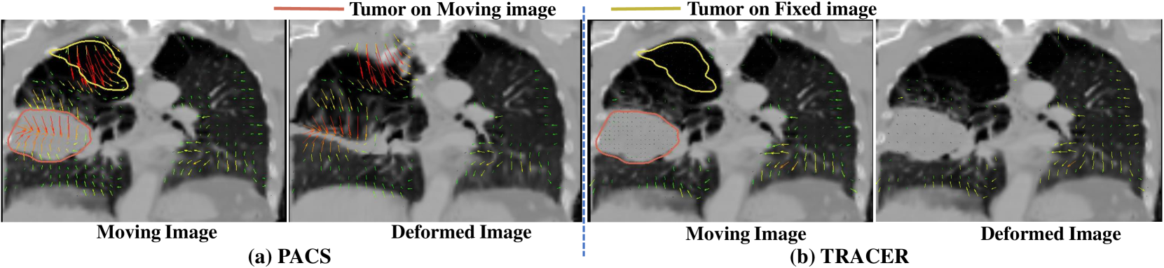
Background: Voxel-based analysis (VBA) for population level radiotherapy (RT) outcomes modeling requires topology preserving inter-patient deformable image registration (DIR) that preserves tumors on moving images while avoiding unrealistic deformations due to tumors occurring on fixed images. Purpose: We developed a tumor-aware recurrent registration (TRACER) deep learning (DL) method and evaluated its suitability for VBA. Methods: TRACER consists of encoder layers implemented with stacked 3D convolutional long short term memory network (3D-CLSTM) followed by decoder and spatial transform layers to compute dense deformation vector field (DVF). Multiple CLSTM steps are used to compute a progressive sequence of deformations. Input conditioning was applied by including tumor segmentations with 3D image pairs as input channels. Bidirectional tumor rigidity, image similarity, and deformation smoothness losses were used to optimize the network in an unsupervised manner. TRACER and multiple DL methods were trained with 204 3D CT image pairs from patients with lung cancers (LC) and evaluated using (a) Dataset I (N = 308 pairs) with DL segmented LCs, (b) Dataset II (N = 765 pairs) with manually delineated LCs, and (c) Dataset III with 42 LC patients treated with RT. Results: TRACER accurately aligned normal tissues. It best preserved tumors, blackindicated by the smallest tumor volume difference of 0.24%, 0.40%, and 0.13 % and mean square error in CT intensities of 0.005, 0.005, 0.004, computed between original and resampled moving image tumors, for Datasets I, II, and III, respectively. It resulted in the smallest planned RT tumor dose difference computed between original and resampled moving images of 0.01 Gy and 0.013 Gy when using a female and a male reference.
Read more9/19/2024

0
Gaussian Representation for Deformable Image Registration
Jihe Li, Fabian Zhang, Xia Li, Tianhao Zhang, Ye Zhang, Joachim Buhmann
Deformable image registration (DIR) is a fundamental task in radiotherapy, with existing methods often struggling to balance computational efficiency, registration accuracy, and speed effectively. We introduce a novel DIR approach employing parametric 3D Gaussian control points achieving a better tradeoff. It provides an explicit and flexible representation for spatial deformation fields between 3D volumetric medical images, producing a displacement vector field (DVF) across all volumetric positions. The movement of individual voxels is derived using linear blend skinning (LBS) through localized interpolation of transformations associated with neighboring Gaussians. This interpolation strategy not only simplifies the determination of voxel motions but also acts as an effective regularization technique. Our approach incorporates a unified optimization process through backpropagation, enabling iterative learning of both the parameters of the 3D Gaussians and their transformations. Additionally, the density of Gaussians is adjusted adaptively during the learning phase to accommodate varying degrees of motion complexity. We validated our approach on the 4D-CT lung DIR-Lab and cardiac ACDC datasets, achieving an average target registration error (TRE) of 1.06 mm within a much-improved processing time of 2.43 seconds for the DIR-Lab dataset over existing methods, demonstrating significant advancements in both accuracy and efficiency.
Read more6/6/2024
🖼️
0
Diffeomorphic Transformer-based Abdomen MRI-CT Deformable Image Registration
Yang Lei, Luke A. Matkovic, Justin Roper, Tonghe Wang, Jun Zhou, Beth Ghavidel, Mark McDonald, Pretesh Patel, Xiaofeng Yang
This paper aims to create a deep learning framework that can estimate the deformation vector field (DVF) for directly registering abdominal MRI-CT images. The proposed method assumed a diffeomorphic deformation. By using topology-preserved deformation features extracted from the probabilistic diffeomorphic registration model, abdominal motion can be accurately obtained and utilized for DVF estimation. The model integrated Swin transformers, which have demonstrated superior performance in motion tracking, into the convolutional neural network (CNN) for deformation feature extraction. The model was optimized using a cross-modality image similarity loss and a surface matching loss. To compute the image loss, a modality-independent neighborhood descriptor (MIND) was used between the deformed MRI and CT images. The surface matching loss was determined by measuring the distance between the warped coordinates of the surfaces of contoured structures on the MRI and CT images. The deformed MRI image was assessed against the CT image using the target registration error (TRE), Dice similarity coefficient (DSC), and mean surface distance (MSD) between the deformed contours of the MRI image and manual contours of the CT image. When compared to only rigid registration, DIR with the proposed method resulted in an increase of the mean DSC values of the liver and portal vein from 0.850 and 0.628 to 0.903 and 0.763, a decrease of the mean MSD of the liver from 7.216 mm to 3.232 mm, and a decrease of the TRE from 26.238 mm to 8.492 mm. The proposed deformable image registration method based on a diffeomorphic transformer provides an effective and efficient way to generate an accurate DVF from an MRI-CT image pair of the abdomen. It could be utilized in the current treatment planning workflow for liver radiotherapy.
Read more5/7/2024

0
Preserving Tumor Volumes for Unsupervised Medical Image Registration
Qihua Dong, Hao Du, Ying Song, Yan Xu, Jing Liao
Medical image registration is a critical task that estimates the spatial correspondence between pairs of images. However, current traditional and deep-learning-based methods rely on similarity measures to generate a deforming field, which often results in disproportionate volume changes in dissimilar regions, especially in tumor regions. These changes can significantly alter the tumor size and underlying anatomy, which limits the practical use of image registration in clinical diagnosis. To address this issue, we have formulated image registration with tumors as a constraint problem that preserves tumor volumes while maximizing image similarity in other normal regions. Our proposed strategy involves a two-stage process. In the first stage, we use similarity-based registration to identify potential tumor regions by their volume change, generating a soft tumor mask accordingly. In the second stage, we propose a volume-preserving registration with a novel adaptive volume-preserving loss that penalizes the change in size adaptively based on the masks calculated from the previous stage. Our approach balances image similarity and volume preservation in different regions, i.e., normal and tumor regions, by using soft tumor masks to adjust the imposition of volume-preserving loss on each one. This ensures that the tumor volume is preserved during the registration process. We have evaluated our strategy on various datasets and network architectures, demonstrating that our method successfully preserves the tumor volume while achieving comparable registration results with state-of-the-art methods. Our codes is available at: url{https://dddraxxx.github.io/Volume-Preserving-Registration/}.
Read more5/13/2024