WEEP: A method for spatial interpretation of weakly supervised CNN models in computational pathology

0
👨🏫
Sign in to get full access
Overview
- Deep learning can be used to analyze high-resolution histopathology whole-slide images (WSI)
- Weakly supervised learning is commonly applied when labels only exist at the patient or WSI level (e.g., patient outcomes or histological grading)
- There is a need for improved spatial interpretability of predictions from these models
- The paper proposes a novel method called WEEP to provide a principled way to identify the spatial area of a WSI required for a particular prediction
Plain English Explanation
Deep learning is a powerful technique that can be used to analyze highly detailed medical images, such as those used in computational pathology for diagnosing diseases like breast cancer. In some cases, the labels or information about these images only exist at a high level, like the overall patient outcome or the histological grade of the tissue sample.
The WEEP method proposed in this paper provides a way to better understand which specific regions of the medical image are most important for the deep learning model's predictions. This can be useful for both research purposes, to gain insights into the disease processes, and for diagnostic applications, to help pathologists interpret the model's decisions.
Imagine you have a high-resolution image of a tissue sample, and a deep learning model can predict whether the patient has a good or bad outcome based on that image. The WEEP method allows you to identify the specific areas of the image that the model is focusing on to make that prediction. This could help researchers understand what features of the tissue are most indicative of the patient's outcome, and it could also help doctors trust the model's predictions by showing them the evidence the model is using.
Technical Explanation
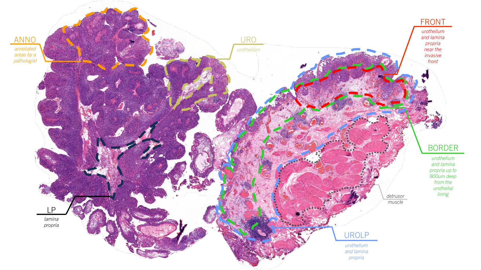
The paper proposes a novel method called the WSI rEgion sElection aPproach (WEEP), which provides a principled yet straightforward way to establish the spatial area of a WSI that is required for assigning a particular prediction label.
The authors demonstrate WEEP on a binary classification task in the domain of breast cancer computational pathology. Weakly supervised learning is typically applied in this context, where labels only exist at the patient or WSI level (e.g., patient outcomes or histological grading), rather than at the level of individual image tiles.
WEEP is designed to offer improved spatial interpretability of the model's predictions by identifying the critical regions of the WSI that contribute most to the classification decision. The method is easy to implement, directly connected to the model-based decision process, and provides information relevant to both research and diagnostic applications.
Critical Analysis
The paper acknowledges that weakly supervised learning approaches, while useful for whole-slide image classification tasks, can lack spatial interpretability. The proposed WEEP method is a step towards addressing this limitation, but the authors note that further research is needed to fully understand the strengths and weaknesses of the approach.
One potential concern is that the WEEP method, while providing a principled way to identify critical regions, may not capture all the nuanced spatial relationships and contextual information that human experts use when interpreting histopathology images. Additional research could explore ways to better incorporate such expert-level understanding into the model interpretation process.
Furthermore, the paper only demonstrates WEEP on a binary classification task in breast cancer computational pathology. Its generalizability to other medical imaging domains and more complex prediction tasks, such as segmentation and classification of specific tumor regions, remains to be evaluated.
Conclusion
The WEEP method proposed in this paper represents a promising approach for improving the spatial interpretability of deep learning models used in computational pathology and other medical imaging applications. By identifying the critical regions of whole-slide images that contribute most to a model's predictions, WEEP can provide valuable insights for both research and diagnostic purposes.
As deep learning continues to automate the prediction of patient outcomes and other clinically relevant factors, methods like WEEP will become increasingly important for ensuring the interpretability and trustworthiness of these powerful AI-driven tools in the medical field.
This summary was produced with help from an AI and may contain inaccuracies - check out the links to read the original source documents!
Related Papers
👨🏫
0
WEEP: A method for spatial interpretation of weakly supervised CNN models in computational pathology
Abhinav Sharma, Bojing Liu, Mattias Rantalainen
Deep learning enables the modelling of high-resolution histopathology whole-slide images (WSI). Weakly supervised learning of tile-level data is typically applied for tasks where labels only exist on the patient or WSI level (e.g. patient outcomes or histological grading). In this context, there is a need for improved spatial interpretability of predictions from such models. We propose a novel method, Wsi rEgion sElection aPproach (WEEP), for model interpretation. It provides a principled yet straightforward way to establish the spatial area of WSI required for assigning a particular prediction label. We demonstrate WEEP on a binary classification task in the area of breast cancer computational pathology. WEEP is easy to implement, is directly connected to the model-based decision process, and offers information relevant to both research and diagnostic applications.
Read more4/9/2024
0
Self-Contrastive Weakly Supervised Learning Framework for Prognostic Prediction Using Whole Slide Images
Saul Fuster, Farbod Khoraminia, Julio Silva-Rodr'iguez, Umay Kiraz, Geert J. L. H. van Leenders, Trygve Eftest{o}l, Valery Naranjo, Emiel A. M. Janssen, Tahlita C. M. Zuiverloon, Kjersti Engan
We present a pioneering investigation into the application of deep learning techniques to analyze histopathological images for addressing the substantial challenge of automated prognostic prediction. Prognostic prediction poses a unique challenge as the ground truth labels are inherently weak, and the model must anticipate future events that are not directly observable in the image. To address this challenge, we propose a novel three-part framework comprising of a convolutional network based tissue segmentation algorithm for region of interest delineation, a contrastive learning module for feature extraction, and a nested multiple instance learning classification module. Our study explores the significance of various regions of interest within the histopathological slides and exploits diverse learning scenarios. The pipeline is initially validated on artificially generated data and a simpler diagnostic task. Transitioning to prognostic prediction, tasks become more challenging. Employing bladder cancer as use case, our best models yield an AUC of 0.721 and 0.678 for recurrence and treatment outcome prediction respectively.
Read more5/27/2024
🖼️
0
Whole Slide Image Survival Analysis Using Histopathological Feature Extractors
Kleanthis Marios Papadopoulos
The abundance of information present in Whole Slide Images (WSIs) makes them useful for prognostic evaluation. A large number of models utilizing a pretrained ResNet backbone have been released and employ various feature aggregation techniques, primarily based on Multiple Instance Learning (MIL). By leveraging the recently released UNI feature extractor, existing models can be adapted to achieve higher accuracy, which paves the way for more robust prognostic tools in digital pathology.
Read more5/29/2024

0
An efficient framework based on large foundation model for cervical cytopathology whole slide image screening
Jialong Huang, Gaojie Li, Shichao Kan, Jianfeng Liu, Yixiong Liang
Current cervical cytopathology whole slide image (WSI) screening primarily relies on detection-based approaches, which are limited in performance due to the expense and time-consuming annotation process. Multiple Instance Learning (MIL), a weakly supervised approach that relies solely on bag-level labels, can effectively alleviate these challenges. Nonetheless, MIL commonly employs frozen pretrained models or self-supervised learning for feature extraction, which suffers from low efficacy or inefficiency. In this paper, we propose an efficient framework for cervical cytopathology WSI classification using only WSI-level labels through unsupervised and weakly supervised learning. Given the sparse and dispersed nature of abnormal cells within cytopathological WSIs, we propose a strategy that leverages the pretrained foundation model to filter the top$k$ high-risk patches. Subsequently, we suggest parameter-efficient fine-tuning (PEFT) of a large foundation model using contrastive learning on the filtered patches to enhance its representation ability for task-specific signals. By training only the added linear adapters, we enhance the learning of patch-level features with substantially reduced time and memory consumption. Experiments conducted on the CSD and FNAC 2019 datasets demonstrate that the proposed method enhances the performance of various MIL methods and achieves state-of-the-art (SOTA) performance. The code and trained models are publicly available at https://github.com/CVIU-CSU/TCT-InfoNCE.
Read more7/17/2024