Whole Slide Image Classification of Salivary Gland Tumours

0

Sign in to get full access
Overview
- This paper presents a deep learning approach for classifying salivary gland tumors from whole slide images.
- The method uses multiple instance learning to handle the large size and complex structure of whole slide images.
- The model achieves state-of-the-art performance on a dataset of salivary gland histopathology slides.
Plain English Explanation
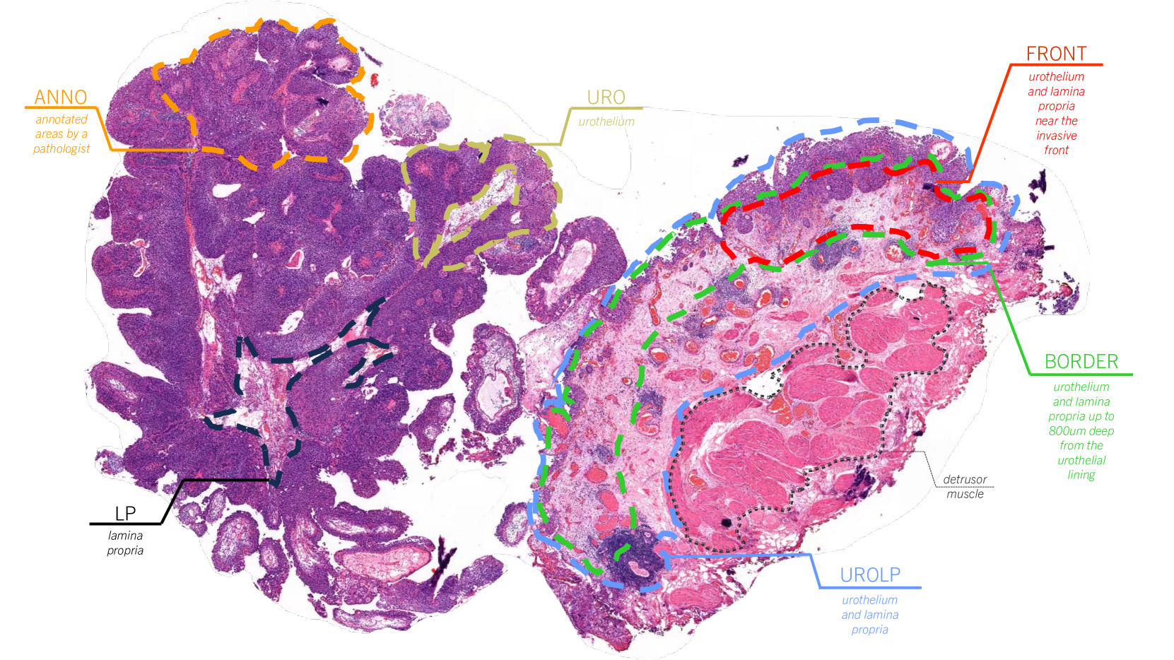
Whole slide images are high-resolution digital scans of entire tissue samples. These images can contain a lot of detailed information, but they can also be very large and complex to analyze. This paper describes a way to use machine learning to automatically classify different types of salivary gland tumors from whole slide images.
The key idea is to use a technique called "multiple instance learning." Rather than trying to classify the entire slide at once, the model breaks the slide down into smaller regions and analyzes each region independently. It then combines the information from all the regions to make an overall classification. This helps the model handle the complexity of the whole slide image.
The researchers trained and tested their model on a dataset of salivary gland histopathology slides. They found that their approach achieved state-of-the-art performance, meaning it was better at classifying the tumor types than previous methods. This suggests that this type of multiple instance learning approach could be useful for analyzing other types of complex medical images as well.
Technical Explanation
The paper presents a multiple instance learning approach for classifying salivary gland tumors from whole slide images. The key steps are:
-
Preprocessing: The whole slide images are divided into small patches, which are then encoded using a pretrained convolutional neural network.
-
Multiple Instance Learning: Instead of classifying the entire slide at once, the model treats each patch as an individual "instance" and learns to classify the slide based on the collective information from all the patches. This allows the model to handle the large size and heterogeneous content of the whole slide images.
-
Weakly Supervised Training: Since the ground truth labels are provided at the slide level rather than the patch level, the model is trained in a weakly supervised manner, relying on the multiple instance learning framework to learn discriminative patch-level features.
-
Evaluation: The trained model is evaluated on a held-out test set of salivary gland histopathology slides. The results show that the multiple instance learning approach outperforms previous methods for this task.
Critical Analysis
The paper provides a thorough technical explanation of the proposed multiple instance learning approach and its application to whole slide image classification of salivary gland tumors. Some potential limitations and areas for further research include:
-
The dataset used in the study, while sizable, may not capture the full diversity of salivary gland tumors seen in clinical practice. Evaluating the model's performance on a larger and more representative dataset could help assess its real-world applicability.
-
The paper does not delve into the interpretability of the model's predictions. Understanding which image regions or features the model is focusing on to make its decisions could provide valuable insights to pathologists and clinicians.
-
While the multiple instance learning framework is well-suited for handling the complexity of whole slide images, exploring other architectural choices or training strategies could potentially further improve the model's performance.
-
The paper does not discuss the computational efficiency or inference time of the proposed approach, which could be an important consideration for real-time clinical deployment.
Overall, the paper presents a promising approach for automating the classification of salivary gland tumors from whole slide images, but additional research may be needed to fully understand the limitations and practical implications of this work.
Conclusion
This paper demonstrates the effectiveness of a multiple instance learning approach for classifying salivary gland tumors from whole slide histopathology images. By breaking down the large and complex whole slide images into smaller, manageable patches and learning to classify the slides based on the collective information from these patches, the model was able to achieve state-of-the-art performance.
The ability to accurately classify salivary gland tumors from digital slides could have significant implications for clinical practice, potentially streamlining the diagnostic process and improving patient outcomes. While the current study is a promising step forward, further research is needed to fully understand the limitations and practical applications of this approach.
This summary was produced with help from an AI and may contain inaccuracies - check out the links to read the original source documents!
Related Papers
0
Whole Slide Image Classification of Salivary Gland Tumours
John Charlton, Ibrahim Alsanie, Syed Ali Khurram
This work shows promising results using multiple instance learning on salivary gland tumours in classifying cancers on whole slide images. Utilising CTransPath as a patch-level feature extractor and CLAM as a feature aggregator, an F1 score of over 0.88 and AUROC of 0.92 are obtained for detecting cancer in whole slide images.
Read more8/23/2024
🖼️
0
Whole Slide Image Survival Analysis Using Histopathological Feature Extractors
Kleanthis Marios Papadopoulos
The abundance of information present in Whole Slide Images (WSIs) makes them useful for prognostic evaluation. A large number of models utilizing a pretrained ResNet backbone have been released and employ various feature aggregation techniques, primarily based on Multiple Instance Learning (MIL). By leveraging the recently released UNI feature extractor, existing models can be adapted to achieve higher accuracy, which paves the way for more robust prognostic tools in digital pathology.
Read more5/29/2024
0
Self-Contrastive Weakly Supervised Learning Framework for Prognostic Prediction Using Whole Slide Images
Saul Fuster, Farbod Khoraminia, Julio Silva-Rodr'iguez, Umay Kiraz, Geert J. L. H. van Leenders, Trygve Eftest{o}l, Valery Naranjo, Emiel A. M. Janssen, Tahlita C. M. Zuiverloon, Kjersti Engan
We present a pioneering investigation into the application of deep learning techniques to analyze histopathological images for addressing the substantial challenge of automated prognostic prediction. Prognostic prediction poses a unique challenge as the ground truth labels are inherently weak, and the model must anticipate future events that are not directly observable in the image. To address this challenge, we propose a novel three-part framework comprising of a convolutional network based tissue segmentation algorithm for region of interest delineation, a contrastive learning module for feature extraction, and a nested multiple instance learning classification module. Our study explores the significance of various regions of interest within the histopathological slides and exploits diverse learning scenarios. The pipeline is initially validated on artificially generated data and a simpler diagnostic task. Transitioning to prognostic prediction, tasks become more challenging. Employing bladder cancer as use case, our best models yield an AUC of 0.721 and 0.678 for recurrence and treatment outcome prediction respectively.
Read more5/27/2024

0
Finding Regions of Interest in Whole Slide Images Using Multiple Instance Learning
Martim Afonso, Praphulla M. S. Bhawsar, Monjoy Saha, Jonas S. Almeida, Arlindo L. Oliveira
Whole Slide Images (WSI), obtained by high-resolution digital scanning of microscope slides at multiple scales, are the cornerstone of modern Digital Pathology. However, they represent a particular challenge to AI-based/AI-mediated analysis because pathology labeling is typically done at slide-level, instead of tile-level. It is not just that medical diagnostics is recorded at the specimen level, the detection of oncogene mutation is also experimentally obtained, and recorded by initiatives like The Cancer Genome Atlas (TCGA), at the slide level. This configures a dual challenge: a) accurately predicting the overall cancer phenotype and b) finding out what cellular morphologies are associated with it at the tile level. To address these challenges, a weakly supervised Multiple Instance Learning (MIL) approach was explored for two prevalent cancer types, Invasive Breast Carcinoma (TCGA-BRCA) and Lung Squamous Cell Carcinoma (TCGA-LUSC). This approach was explored for tumor detection at low magnification levels and TP53 mutations at various levels. Our results show that a novel additive implementation of MIL matched the performance of reference implementation (AUC 0.96), and was only slightly outperformed by Attention MIL (AUC 0.97). More interestingly from the perspective of the molecular pathologist, these different AI architectures identify distinct sensitivities to morphological features (through the detection of Regions of Interest, RoI) at different amplification levels. Tellingly, TP53 mutation was most sensitive to features at the higher applications where cellular morphology is resolved.
Read more4/12/2024