Inpainting Pathology in Lumbar Spine MRI with Latent Diffusion
2406.02477
0
0

Abstract
Data driven models for automated diagnosis in radiology suffer from insufficient and imbalanced datasets due to low representation of pathology in a population and the cost of expert annotations. Datasets can be bolstered through data augmentation. However, even when utilizing a full suite of transformations during model training, typical data augmentations do not address variations in human anatomy. An alternative direction is to synthesize data using generative models, which can potentially craft datasets with specific attributes. While this holds promise, commonly used generative models such as Generative Adversarial Networks may inadvertently produce anatomically inaccurate features. On the other hand, diffusion models, which offer greater stability, tend to memorize training data, raising concerns about privacy and generative diversity. Alternatively, inpainting has the potential to augment data through directly inserting pathology in medical images. However, this approach introduces a new challenge: accurately merging the generated pathological features with the surrounding anatomical context. While inpainting is a well established method for addressing simple lesions, its application to pathologies that involve complex structural changes remains relatively unexplored. We propose an efficient method for inpainting pathological features onto healthy anatomy in MRI through voxelwise noise scheduling in a latent diffusion model. We evaluate the method's ability to insert disc herniation and central canal stenosis in lumbar spine sagittal T2 MRI, and it achieves superior Frechet Inception Distance compared to state-of-the-art methods.
Create account to get full access
Overview
- This paper presents a method for inpainting pathology in lumbar spine MRI scans using a latent diffusion model.
- The approach aims to restore missing or corrupted regions in medical images, which can be critical for accurate diagnosis and treatment.
- The authors demonstrate the effectiveness of their method on a dataset of lumbar spine MRI scans with simulated pathological regions.
Plain English Explanation
Medical imaging, like MRI scans of the spine, can sometimes have missing or distorted areas due to various issues during the scanning process. This can make it difficult for doctors to properly diagnose and treat patients. The researchers in this paper have developed a new technique using latent diffusion models to "inpaint" or fill in these problematic regions in spine MRI scans.
The key idea is to train an AI model that can look at the surrounding healthy tissue in the MRI scan and intelligently "guess" what the missing or distorted area should look like, based on patterns in the data. This allows the doctors to get a clearer, more complete picture of the patient's spine without having to repeat the entire MRI process.
The researchers tested their method on a dataset of lumbar spine MRI scans where they intentionally removed or corrupted certain regions to simulate pathological issues. They found that their latent diffusion inpainting approach was able to accurately restore these simulated problem areas, suggesting it could be a valuable tool for real-world medical applications.
Technical Explanation
The paper presents a method for inpainting pathology in lumbar spine MRI scans using a latent diffusion model. Latent diffusion models [1] are a type of generative AI that can be used to synthesize or manipulate high-dimensional data like images.
The authors' approach involves training a latent diffusion model on a dataset of lumbar spine MRI scans. During training, the model learns to map the MRI scans into a lower-dimensional "latent space" representation. The model is then trained to generate plausible latent representations for corrupted or missing regions of the MRI scans, which can be decoded back into the original image space to "inpaint" the problematic areas.
The researchers evaluate their method on a dataset of lumbar spine MRI scans where they have simulated pathological issues by removing or distorting certain regions of the images. They find that their latent diffusion inpainting approach is able to accurately restore these simulated problem areas, outperforming several baseline methods.
Critical Analysis
The paper presents a novel and promising approach for addressing the important problem of inpainting pathology in medical images. The use of latent diffusion models allows the method to effectively "hallucinate" plausible content for missing or corrupted regions, which could be a valuable tool for clinicians.
However, the paper does have some limitations. The experiments are conducted on simulated pathological regions rather than real clinical data, so further testing is needed to demonstrate the approach's effectiveness on genuine patient scans. Additionally, the dataset used is relatively small, which may limit the generalizability of the results.
It would also be interesting to see how this method compares to other diffusion-based inpainting techniques or approaches that leverage anomaly detection for medical image restoration.
Overall, this research represents an important step forward in using generative AI models to address challenges in medical imaging, but further work is needed to fully validate the approach and understand its clinical utility.
Conclusion
This paper introduces a latent diffusion-based method for inpainting pathology in lumbar spine MRI scans. The key innovation is the use of a generative AI model to intelligently "fill in" missing or corrupted regions of medical images, which could significantly aid clinicians in diagnosis and treatment.
While the results on simulated data are promising, further testing on real-world clinical datasets is needed to fully assess the approach's capabilities. Nonetheless, this research represents an important step forward in leveraging advanced machine learning techniques to tackle critical problems in healthcare.
This summary was produced with help from an AI and may contain inaccuracies - check out the links to read the original source documents!
Related Papers
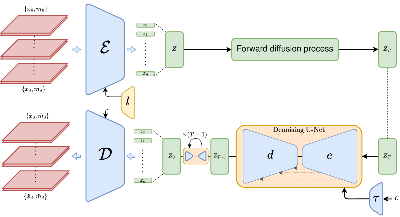
3D MRI Synthesis with Slice-Based Latent Diffusion Models: Improving Tumor Segmentation Tasks in Data-Scarce Regimes
Aghiles Kebaili, J'er^ome Lapuyade-Lahorgue, Pierre Vera, Su Ruan
0
0
Despite the increasing use of deep learning in medical image segmentation, the limited availability of annotated training data remains a major challenge due to the time-consuming data acquisition and privacy regulations. In the context of segmentation tasks, providing both medical images and their corresponding target masks is essential. However, conventional data augmentation approaches mainly focus on image synthesis. In this study, we propose a novel slice-based latent diffusion architecture designed to address the complexities of volumetric data generation in a slice-by-slice fashion. This approach extends the joint distribution modeling of medical images and their associated masks, allowing a simultaneous generation of both under data-scarce regimes. Our approach mitigates the computational complexity and memory expensiveness typically associated with diffusion models. Furthermore, our architecture can be conditioned by tumor characteristics, including size, shape, and relative position, thereby providing a diverse range of tumor variations. Experiments on a segmentation task using the BRATS2022 confirm the effectiveness of the synthesized volumes and masks for data augmentation.
6/11/2024

Taming Latent Diffusion Model for Neural Radiance Field Inpainting
Chieh Hubert Lin, Changil Kim, Jia-Bin Huang, Qinbo Li, Chih-Yao Ma, Johannes Kopf, Ming-Hsuan Yang, Hung-Yu Tseng
0
0
Neural Radiance Field (NeRF) is a representation for 3D reconstruction from multi-view images. Despite some recent work showing preliminary success in editing a reconstructed NeRF with diffusion prior, they remain struggling to synthesize reasonable geometry in completely uncovered regions. One major reason is the high diversity of synthetic contents from the diffusion model, which hinders the radiance field from converging to a crisp and deterministic geometry. Moreover, applying latent diffusion models on real data often yields a textural shift incoherent to the image condition due to auto-encoding errors. These two problems are further reinforced with the use of pixel-distance losses. To address these issues, we propose tempering the diffusion model's stochasticity with per-scene customization and mitigating the textural shift with masked adversarial training. During the analyses, we also found the commonly used pixel and perceptual losses are harmful in the NeRF inpainting task. Through rigorous experiments, our framework yields state-of-the-art NeRF inpainting results on various real-world scenes. Project page: https://hubert0527.github.io/MALD-NeRF
4/16/2024
Diffusion-based image inpainting with internal learning
Nicolas Cherel, Andr'es Almansa, Yann Gousseau, Alasdair Newson
0
0
Diffusion models are now the undisputed state-of-the-art for image generation and image restoration. However, they require large amounts of computational power for training and inference. In this paper, we propose lightweight diffusion models for image inpainting that can be trained on a single image, or a few images. We show that our approach competes with large state-of-the-art models in specific cases. We also show that training a model on a single image is particularly relevant for image acquisition modality that differ from the RGB images of standard learning databases. We show results in three different contexts: texture images, line drawing images, and materials BRDF, for which we achieve state-of-the-art results in terms of realism, with a computational load that is greatly reduced compared to concurrent methods.
6/7/2024
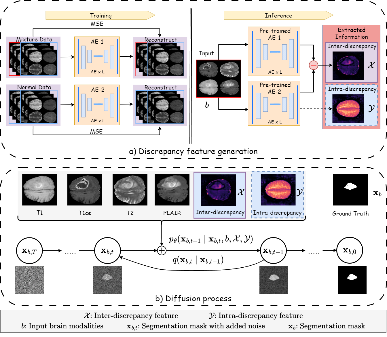
Discrepancy-based Diffusion Models for Lesion Detection in Brain MRI
Keqiang Fan, Xiaohao Cai, Mahesan Niranjan
0
0
Diffusion probabilistic models (DPMs) have exhibited significant effectiveness in computer vision tasks, particularly in image generation. However, their notable performance heavily relies on labelled datasets, which limits their application in medical images due to the associated high-cost annotations. Current DPM-related methods for lesion detection in medical imaging, which can be categorized into two distinct approaches, primarily rely on image-level annotations. The first approach, based on anomaly detection, involves learning reference healthy brain representations and identifying anomalies based on the difference in inference results. In contrast, the second approach, resembling a segmentation task, employs only the original brain multi-modalities as prior information for generating pixel-level annotations. In this paper, our proposed model - discrepancy distribution medical diffusion (DDMD) - for lesion detection in brain MRI introduces a novel framework by incorporating distinctive discrepancy features, deviating from the conventional direct reliance on image-level annotations or the original brain modalities. In our method, the inconsistency in image-level annotations is translated into distribution discrepancies among heterogeneous samples while preserving information within homogeneous samples. This property retains pixel-wise uncertainty and facilitates an implicit ensemble of segmentation, ultimately enhancing the overall detection performance. Thorough experiments conducted on the BRATS2020 benchmark dataset containing multimodal MRI scans for brain tumour detection demonstrate the great performance of our approach in comparison to state-of-the-art methods.
5/9/2024