Cardiac Copilot: Automatic Probe Guidance for Echocardiography with World Model

0

Sign in to get full access
Overview
- This paper presents "Cardiac Copilot," a system that uses a world model to automatically guide the probe during echocardiography, a common medical imaging technique for the heart.
- The system aims to help sonographers (medical professionals who perform echocardiography) obtain high-quality images by providing real-time probe guidance and navigation.
- The key innovation is the use of a learned world model that maps the patient's cardiac anatomy to the probe's position and orientation, allowing the system to recommend optimal probe movements.
Plain English Explanation
<a href="https://aimodels.fyi/papers/arxiv/epicardium-prompt-guided-real-time-cardiac-ultrasound">Echocardiography</a> is a widely used medical imaging technique that uses sound waves to create images of the heart. It helps doctors diagnose and monitor heart conditions. However, getting high-quality images can be challenging, as the sonographer needs to carefully maneuver the ultrasound probe around the patient's chest to capture the right views.
The "Cardiac Copilot" system aims to assist sonographers by automatically guiding the probe during the echocardiography procedure. It does this by using a "world model" - a computer simulation of the patient's cardiac anatomy and how it relates to the probe's position and orientation. This world model allows the system to continuously track the probe's location and recommend the best movements to capture the desired views of the heart.
<a href="https://aimodels.fyi/papers/arxiv/goal-conditioned-reinforcement-learning-ultrasound-navigation-guidance">For example</a>, if the sonographer is trying to get a view of the heart's left ventricle, the system can analyze the probe's current position and suggest small adjustments to help the sonographer obtain the optimal view. By providing this real-time guidance, the system helps the sonographer work more efficiently and capture high-quality images more consistently.
Technical Explanation
The key innovation in the "Cardiac Copilot" system is the use of a learned <a href="https://aimodels.fyi/papers/arxiv/prompt-driven-universal-model-view-agnostic-echocardiography">world model</a> that maps the patient's cardiac anatomy to the probe's position and orientation. This world model is trained on a large dataset of echocardiography images and probe tracking data, allowing it to learn the complex relationships between the probe's movements and the resulting images.
During the echocardiography procedure, the system uses this world model to continuously track the probe's location and orientation relative to the patient's heart. It then analyzes the current probe position and suggests optimal movements to capture the desired view of the heart, such as adjusting the probe's angle or depth.
<a href="https://aimodels.fyi/papers/arxiv/guiding-last-centimeter-novel-anatomy-aware-probe">The system's recommendations are based on a deep understanding of cardiac anatomy and the probe's kinematics</a>, allowing it to provide precise and tailored guidance to the sonographer. This helps the sonographer obtain high-quality images more efficiently, ultimately improving the diagnostic process and patient care.
Critical Analysis
The "Cardiac Copilot" system represents a significant advancement in the field of echocardiography automation and guidance. By leveraging a learned world model, the system can provide real-time, personalized probe guidance that adapts to the unique anatomy of each patient.
However, the paper does not address the potential challenges of integrating such a system into clinical workflows. Sonographers may be hesitant to relinquish full control of the probe, and the system's recommendations may not always align with the sonographer's expertise and intuition.
Additionally, the <a href="https://aimodels.fyi/papers/arxiv/echotracker-advancing-myocardial-point-tracking-echocardiography">accuracy and reliability of the world model</a> are crucial to the system's performance. Further research is needed to validate the model's robustness across a diverse patient population and various cardiac conditions.
Conclusion
The "Cardiac Copilot" system presents a promising approach to improving the efficiency and consistency of echocardiography procedures. By automating probe guidance using a learned world model, the system has the potential to help sonographers capture high-quality images more easily, leading to better diagnostic capabilities and patient outcomes.
As with any new technology, careful integration with existing clinical practices and continued validation will be necessary to ensure the system's widespread adoption and long-term success. However, the concepts and techniques demonstrated in this paper represent an important step forward in the field of medical imaging automation and guidance.
This summary was produced with help from an AI and may contain inaccuracies - check out the links to read the original source documents!
Related Papers
0
Cardiac Copilot: Automatic Probe Guidance for Echocardiography with World Model
Haojun Jiang, Zhenguo Sun, Ning Jia, Meng Li, Yu Sun, Shaqi Luo, Shiji Song, Gao Huang
Echocardiography is the only technique capable of real-time imaging of the heart and is vital for diagnosing the majority of cardiac diseases. However, there is a severe shortage of experienced cardiac sonographers, due to the heart's complex structure and significant operational challenges. To mitigate this situation, we present a Cardiac Copilot system capable of providing real-time probe movement guidance to assist less experienced sonographers in conducting freehand echocardiography. This system can enable non-experts, especially in primary departments and medically underserved areas, to perform cardiac ultrasound examinations, potentially improving global healthcare delivery. The core innovation lies in proposing a data-driven world model, named Cardiac Dreamer, for representing cardiac spatial structures. This world model can provide structure features of any cardiac planes around the current probe position in the latent space, serving as an precise navigation map for autonomous plane localization. We train our model with real-world ultrasound data and corresponding probe motion from 110 routine clinical scans with 151K sample pairs by three certified sonographers. Evaluations on three standard planes with 37K sample pairs demonstrate that the world model can reduce navigation errors by up to 33% and exhibit more stable performance.
Read more6/21/2024

0
Structure-aware World Model for Probe Guidance via Large-scale Self-supervised Pre-train
Haojun Jiang, Meng Li, Zhenguo Sun, Ning Jia, Yu Sun, Shaqi Luo, Shiji Song, Gao Huang
The complex structure of the heart leads to significant challenges in echocardiography, especially in acquisition cardiac ultrasound images. Successful echocardiography requires a thorough understanding of the structures on the two-dimensional plane and the spatial relationships between planes in three-dimensional space. In this paper, we innovatively propose a large-scale self-supervised pre-training method to acquire a cardiac structure-aware world model. The core innovation lies in constructing a self-supervised task that requires structural inference by predicting masked structures on a 2D plane and imagining another plane based on pose transformation in 3D space. To support large-scale pre-training, we collected over 1.36 million echocardiograms from ten standard views, along with their 3D spatial poses. In the downstream probe guidance task, we demonstrate that our pre-trained model consistently reduces guidance errors across the ten most common standard views on the test set with 0.29 million samples from 74 routine clinical scans, indicating that structure-aware pre-training benefits the scanning.
Read more7/22/2024

0
Sequence-aware Pre-training for Echocardiography Probe Guidance
Haojun Jiang, Zhenguo Sun, Yu Sun, Ning Jia, Meng Li, Shaqi Luo, Shiji Song, Gao Huang
Cardiac ultrasound probe guidance aims to help novices adjust the 6-DOF probe pose to obtain high-quality sectional images. Cardiac ultrasound faces two major challenges: (1) the inherently complex structure of the heart, and (2) significant individual variations. Previous works have only learned the population-averaged 2D and 3D structures of the heart rather than personalized cardiac structural features, leading to a performance bottleneck. Clinically, we observed that sonographers adjust their understanding of a patient's cardiac structure based on prior scanning sequences, thereby modifying their scanning strategies. Inspired by this, we propose a sequence-aware self-supervised pre-training method. Specifically, our approach learns personalized 2D and 3D cardiac structural features by predicting the masked-out images and actions in a scanning sequence. We hypothesize that if the model can predict the missing content it has acquired a good understanding of the personalized cardiac structure. In the downstream probe guidance task, we also introduced a sequence modeling approach that models individual cardiac structural information based on the images and actions from historical scan data, enabling more accurate navigation decisions. Experiments on a large-scale dataset with 1.36 million samples demonstrated that our proposed sequence-aware paradigm can significantly reduce navigation errors, with translation errors decreasing by 15.90% to 36.87% and rotation errors decreasing by 11.13% to 20.77%, compared to state-of-the-art methods.
Read more8/28/2024
0
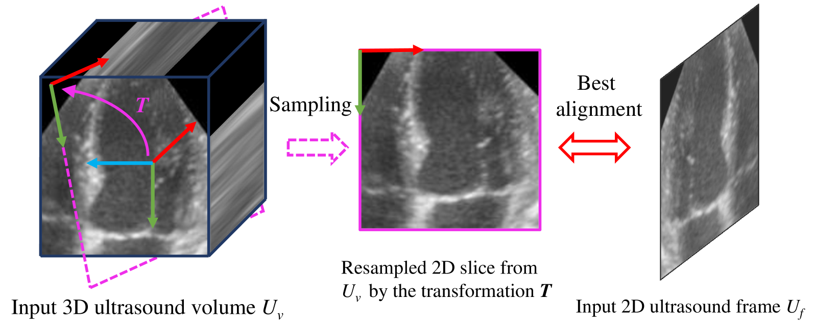
Epicardium Prompt-guided Real-time Cardiac Ultrasound Frame-to-volume Registration
Long Lei, Jun Zhou, Jialun Pei, Baoliang Zhao, Yueming Jin, Yuen-Chun Jeremy Teoh, Jing Qin, Pheng-Ann Heng
A comprehensive guidance view for cardiac interventional surgery can be provided by the real-time fusion of the intraoperative 2D images and preoperative 3D volume based on the ultrasound frame-to-volume registration. However, cardiac ultrasound images are characterized by a low signal-to-noise ratio and small differences between adjacent frames, coupled with significant dimension variations between 2D frames and 3D volumes to be registered, resulting in real-time and accurate cardiac ultrasound frame-to-volume registration being a very challenging task. This paper introduces a lightweight end-to-end Cardiac Ultrasound frame-to-volume Registration network, termed CU-Reg. Specifically, the proposed model leverages epicardium prompt-guided anatomical clues to reinforce the interaction of 2D sparse and 3D dense features, followed by a voxel-wise local-global aggregation of enhanced features, thereby boosting the cross-dimensional matching effectiveness of low-quality ultrasound modalities. We further embed an inter-frame discriminative regularization term within the hybrid supervised learning to increase the distinction between adjacent slices in the same ultrasound volume to ensure registration stability. Experimental results on the reprocessed CAMUS dataset demonstrate that our CU-Reg surpasses existing methods in terms of registration accuracy and efficiency, meeting the guidance requirements of clinical cardiac interventional surgery.
Read more7/1/2024