Cherenkov Imaged Bio-morphological Features Verify Patient Positioning with Deformable Tissue Translocation in Breast Radiotherapy

0
🤖
Sign in to get full access
Overview
- Accurate patient positioning is crucial for effective radiotherapy treatment.
- This study introduces a novel method to track local tissue deformation using Cherenkov imaging during breast cancer radiotherapy.
- The goal was to develop an algorithm to quantify regional positioning accuracy, focusing on loco-regional deformations.
Plain English Explanation
Radiotherapy is a common cancer treatment that uses high-energy radiation to destroy tumor cells. For this treatment to be effective, it's important that the radiation is precisely delivered to the target area and avoids healthy surrounding tissues as much as possible. Accurate patient positioning is critical to achieve this, as even small errors in positioning can significantly impact the treatment outcome.
This study looked at a new way to track changes in the patient's position and shape of the treated area during radiotherapy. The researchers used a technique called Cherenkov imaging, which detects the faint blue light emitted when the radiation passes through the body. By analyzing these Cherenkov images, the team developed an algorithm to quantify both global shifts in the patient's position as well as more localized deformations in the treated region.
The approach was tested using a phantom (model) of the chest area, where known movements were simulated to assess the accuracy. The researchers then applied the method to Cherenkov images collected from 10 actual breast cancer patients undergoing radiotherapy. This allowed them to measure the variation in patient positioning between treatment sessions (inter-fraction) as well as changes within a single session (intra-fraction).
The results showed that this Cherenkov-based technique was able to detect and quantify these positioning variations, including the local deformations that are difficult to capture with conventional imaging methods. This could help improve the precision of radiotherapy delivery and ultimately lead to better treatment outcomes for patients.
Technical Explanation
The study used a combined rigid and non-rigid registration technique to detect inter- and intra-fractional positioning variations from Cherenkov images. First, a rigid registration was performed to quantify global shifts in the patient's position. Then, a non-rigid registration was used to create a two-dimensional map of the loco-regional deformations.
This approach was validated using an anthropomorphic chest phantom experiment, where known couch translations and respiratory motion were simulated. The method achieved an average accuracy of 0.83 mm for couch translations up to 20 mm.
Analysis of Cherenkov data from 10 breast cancer patients undergoing fractionated radiotherapy showed an inter-fraction setup variation of 3.7 ± 2.4 mm relative to the first fraction. Loco-regional deformations (95th percentile) of up to 3.3 ± 1.9 mm were also detected.
Critical Analysis
The study demonstrates the feasibility of using Cherenkov imaging to quantify both global and local positioning variations during radiotherapy, which is a significant advancement over conventional imaging techniques. However, the sample size of 10 patients is relatively small, and more extensive clinical validation would be necessary to fully establish the reliability and robustness of the method.
Additionally, the study focused on breast cancer radiotherapy, and further research would be needed to determine if the approach can be effectively applied to other cancer types and treatment sites. Potential challenges, such as the influence of skin pigmentation or the presence of surgical scars on Cherenkov image quality, should also be investigated.
Conclusion
This study presents a novel Cherenkov-based approach to quantify global and local positioning variations during radiotherapy. The method demonstrated the ability to detect both inter-fraction setup errors and intra-fraction loco-regional deformations, which are critical factors for precise dose delivery and improved treatment outcomes. While further validation is needed, this work represents an important step towards more accurate and personalized radiotherapy.
This summary was produced with help from an AI and may contain inaccuracies - check out the links to read the original source documents!
Related Papers
🤖
0
Cherenkov Imaged Bio-morphological Features Verify Patient Positioning with Deformable Tissue Translocation in Breast Radiotherapy
Yao Chen, Savannah M. Decker, Petr Bruza, David J. Gladstone, Lesley A. Jarvis, Brian W. Pogue, Kimberley S. Samkoe, Rongxiao Zhang
Accurate patient positioning is critical for precise radiotherapy dose delivery, as positioning errors can significantly affect treatment outcomes. This study introduces a novel method for tracking loco-regional tissue deformation through Cherenkov image analysis during fractionated breast cancer radiotherapy. The primary goal was to develop and test an algorithm for Cherenkov-based regional position accuracy quantification, specifically for loco-regional deformations, which lack ideal quantification methods in radiotherapy. Blood vessel detection and segmentation were developed in Cherenkov images using a tissue phantom with incremental movements, and later applied to images from fractionated whole breast radiotherapy in human patients (n=10). A combined rigid and non-rigid registration technique was used to detect inter- and intra-fractional positioning variations. This approach quantified positioning variations in two parts: a global shift from rigid registration and a two-dimensional variation map of loco-regional deformation from non-rigid registration. The methodology was validated using an anthropomorphic chest phantom experiment, where known treatment couch translations and respiratory motion were simulated to assess inter- and intra-fractional uncertainties, yielding an average accuracy of 0.83 mm for couch translations up to 20 mm. Analysis of clinical Cherenkov data from ten breast cancer patients showed an inter-fraction setup variation of 3.7 plus minus 2.4 mm relative to the first fraction and loco-regional deformations (95th percentile) of up to 3.3 plus minus 1.9 mm. This study presents a Cherenkov-based approach to quantify global and local positioning variations, demonstrating feasibility in addressing loco-regional deformations that conventional imaging techniques fail to capture.
Read more9/10/2024
🤿
0
Robust Real-time Segmentation of Bio-Morphological Features in Human Cherenkov Imaging during Radiotherapy via Deep Learning
Shiru Wang, Yao Chen, Lesley A. Jarvis, Yucheng Tang, David J. Gladstone, Kimberley S. Samkoe, Brian W. Pogue, Petr Bruza, Rongxiao Zhang
Cherenkov imaging enables real-time visualization of megavoltage X-ray or electron beam delivery to the patient during Radiation Therapy (RT). Bio-morphological features, such as vasculature, seen in these images are patient-specific signatures that can be used for verification of positioning and motion management that are essential to precise RT treatment. However until now, no concerted analysis of this biological feature-based tracking was utilized because of the slow speed and accuracy of conventional image processing for feature segmentation. This study demonstrated the first deep learning framework for such an application, achieving video frame rate processing. To address the challenge of limited annotation of these features in Cherenkov images, a transfer learning strategy was applied. A fundus photography dataset including 20,529 patch retina images with ground-truth vessel annotation was used to pre-train a ResNet segmentation framework. Subsequently, a small Cherenkov dataset (1,483 images from 212 treatment fractions of 19 breast cancer patients) with known annotated vasculature masks was used to fine-tune the model for accurate segmentation prediction. This deep learning framework achieved consistent and rapid segmentation of Cherenkov-imaged bio-morphological features on another 19 patients, including subcutaneous veins, scars, and pigmented skin. Average segmentation by the model achieved Dice score of 0.85 and required less than 0.7 milliseconds processing time per instance. The model demonstrated outstanding consistency against input image variances and speed compared to conventional manual segmentation methods, laying the foundation for online segmentation in real-time monitoring in a prospective setting.
Read more9/10/2024
0
New!Tumor aware recurrent inter-patient deformable image registration of computed tomography scans with lung cancer
Jue Jiang, Chloe Min Seo Choi, Maria Thor, Joseph O. Deasy, Harini Veeraraghavan
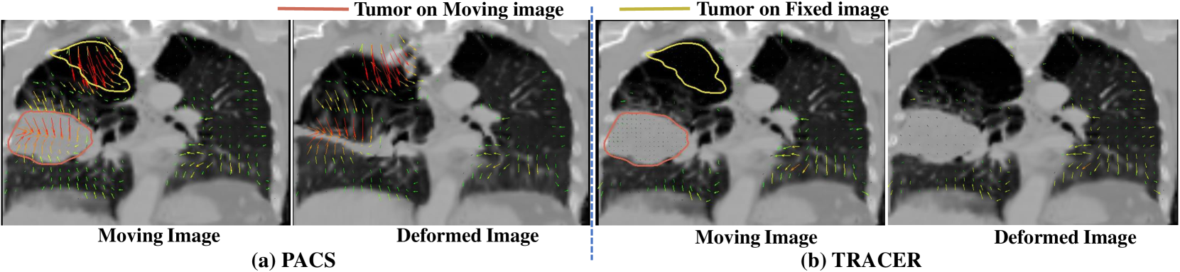
Background: Voxel-based analysis (VBA) for population level radiotherapy (RT) outcomes modeling requires topology preserving inter-patient deformable image registration (DIR) that preserves tumors on moving images while avoiding unrealistic deformations due to tumors occurring on fixed images. Purpose: We developed a tumor-aware recurrent registration (TRACER) deep learning (DL) method and evaluated its suitability for VBA. Methods: TRACER consists of encoder layers implemented with stacked 3D convolutional long short term memory network (3D-CLSTM) followed by decoder and spatial transform layers to compute dense deformation vector field (DVF). Multiple CLSTM steps are used to compute a progressive sequence of deformations. Input conditioning was applied by including tumor segmentations with 3D image pairs as input channels. Bidirectional tumor rigidity, image similarity, and deformation smoothness losses were used to optimize the network in an unsupervised manner. TRACER and multiple DL methods were trained with 204 3D CT image pairs from patients with lung cancers (LC) and evaluated using (a) Dataset I (N = 308 pairs) with DL segmented LCs, (b) Dataset II (N = 765 pairs) with manually delineated LCs, and (c) Dataset III with 42 LC patients treated with RT. Results: TRACER accurately aligned normal tissues. It best preserved tumors, blackindicated by the smallest tumor volume difference of 0.24%, 0.40%, and 0.13 % and mean square error in CT intensities of 0.005, 0.005, 0.004, computed between original and resampled moving image tumors, for Datasets I, II, and III, respectively. It resulted in the smallest planned RT tumor dose difference computed between original and resampled moving images of 0.01 Gy and 0.013 Gy when using a female and a male reference.
Read more9/19/2024
👨🏫
0
Weakly supervised alignment and registration of MR-CT for cervical cancer radiotherapy
Jjahao Zhang, Yin Gu, Deyu Sun, Yuhua Gao, Ming Gao, Ming Cui, Teng Zhang, He Ma
Cervical cancer is one of the leading causes of death in women, and brachytherapy is currently the primary treatment method. However, it is important to precisely define the extent of paracervical tissue invasion to improve cancer diagnosis and treatment options. The fusion of the information characteristics of both computed tomography (CT) and magnetic resonance imaging(MRI) modalities may be useful in achieving a precise outline of the extent of paracervical tissue invasion. Registration is the initial step in information fusion. However, when aligning multimodal images with varying depths, manual alignment is prone to large errors and is time-consuming. Furthermore, the variations in the size of the Region of Interest (ROI) and the shape of multimodal images pose a significant challenge for achieving accurate registration.In this paper, we propose a preliminary spatial alignment algorithm and a weakly supervised multimodal registration network. The spatial position alignment algorithm efficiently utilizes the limited annotation information in the two modal images provided by the doctor to automatically align multimodal images with varying depths. By utilizing aligned multimodal images for weakly supervised registration and incorporating pyramidal features and cost volume to estimate the optical flow, the results indicate that the proposed method outperforms traditional volume rendering alignment methods and registration networks in various evaluation metrics. This demonstrates the effectiveness of our model in multimodal image registration.
Read more5/22/2024